
Can a Portable Wireless Ultrasound Scanner Reliably Evaluate Residual Pericardial Effusion After Cardiac Tamponade?
Yes. Modern portable ultrasound technology enables clinicians to perform rapid, high-quality bedside echocardiography in patients recovering from cardiac tamponade. A wireless ultrasound scanner provides immediate visualization of the pericardial space, allowing physicians to distinguish between clinically insignificant residual pericardial fluid and findings that require urgent intervention.
This clinical case demonstrates how a follow-up cardiac ultrasound examination performed with the Sante Integrale Universal Wi-Fi Mini Ultrasound Scanner supported evidence-based clinical decision-making after successful pericardiocentesis. Rather than relying solely on the presence of residual fluid, management was guided by the patient’s clinical status and echocardiographic findings, consistent with current international recommendations.
The case also highlights how modern portable ultrasound systems have become an integral part of contemporary cardiovascular care, providing physicians with rapid diagnostic information wherever the patient is located—from emergency departments and intensive care units to outpatient clinics and bedside examinations.
Clinical Case
Patient: Male, 56 years old.
Two weeks before the follow-up examination, the patient was admitted to the emergency department with progressive shortness of breath, generalized weakness, dizziness, and reduced exercise tolerance. Symptoms had gradually worsened over several days, eventually limiting even minimal physical activity.
Initial assessment revealed hypotension, sinus tachycardia, elevated jugular venous pressure, and clinical signs suggestive of impaired cardiac filling. Heart sounds were mildly muffled on auscultation. Given the combination of hemodynamic instability and clinical findings, urgent cardiac imaging was immediately performed.
Emergency transthoracic echocardiography demonstrated a large circumferential pericardial effusion with classic echocardiographic features of cardiac tamponade, including right atrial and right ventricular diastolic compression, consistent with life-threatening impairment of cardiac filling.
Urgent pericardiocentesis was performed, evacuating a significant volume of inflammatory pericardial fluid. The patient’s blood pressure improved rapidly after drainage, tachycardia resolved, and symptoms progressively disappeared over the following hours.
Subsequent laboratory evaluation and clinical assessment supported the diagnosis of acute idiopathic, most likely viral, pericarditis complicated by cardiac tamponade. Anti-inflammatory treatment with nonsteroidal anti-inflammatory drugs (NSAIDs) and colchicine was initiated according to current clinical practice guidelines.
Two weeks later, the patient returned for scheduled follow-up evaluation. At this time he reported complete resolution of dyspnea, no chest discomfort, no dizziness, and no exercise limitation. Blood pressure and heart rate were within normal limits, and physical examination showed no evidence of recurrent hemodynamic compromise.
A follow-up echocardiographic examination was performed using the Sante Integrale Universal Wi-Fi Mini Ultrasound Scanner, a portable wireless ultrasound system designed for point-of-care cardiac imaging.
Follow-Up Echocardiographic Examination
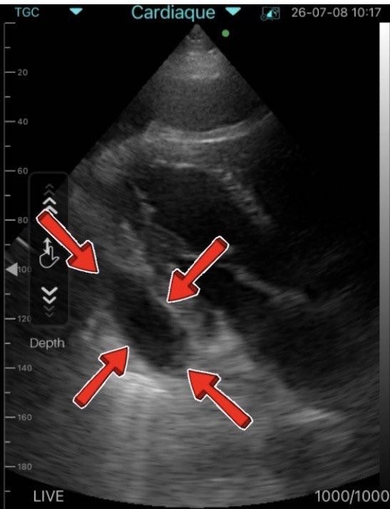
The follow-up examination demonstrated a small localized residual pericardial effusion adjacent to the lateral wall of the left ventricle. Importantly, no echocardiographic evidence of cardiac chamber compression was identified.
The right atrium and right ventricle maintained normal diastolic filling without collapse. Cardiac motion remained preserved, and there were no sonographic signs of recurrent cardiac tamponade. Hemodynamic parameters appeared stable throughout the examination.
From a clinical perspective, the patient remained completely asymptomatic. Blood pressure was within the normal range, heart rate was regular, oxygen saturation was normal, and there were no signs of heart failure or systemic venous congestion.
The echocardiographic findings were fully consistent with an uncomplicated recovery following successful pericardiocentesis.
Clinical Questions
- What is the most likely diagnosis?
- Does this patient require repeat pericardial drainage?
- What is the appropriate management strategy?
Discussion
The most likely diagnosis is acute idiopathic pericarditis, most probably of viral origin, complicated by cardiac tamponade.
Idiopathic and viral pericarditis remain the leading causes of large inflammatory pericardial effusions in countries where tuberculosis is uncommon. In these patients, prompt recognition of tamponade physiology and immediate pericardial drainage are often lifesaving.
The follow-up echocardiogram demonstrates a small residual localized pericardial effusion, a finding frequently observed during recovery after acute pericarditis and successful pericardiocentesis. Residual fluid alone should not be interpreted as treatment failure.
Clinical management should always be based on the overall clinical picture rather than the echocardiographic appearance in isolation.
In this patient:
- clinical symptoms have completely resolved;
- blood pressure and heart rate are stable;
- there is no evidence of recurrent hemodynamic compromise;
- right-sided cardiac chambers are not compressed;
- no echocardiographic signs of tamponade physiology are present.
Therefore, repeat pericardiocentesis is not indicated.
Instead, current international guidelines recommend continuation of anti-inflammatory therapy together with scheduled clinical follow-up and repeat echocardiographic assessment until the pericardial effusion resolves completely or becomes clinically insignificant.
This case illustrates an important educational principle in cardiovascular ultrasound:
A small residual pericardial effusion after successful treatment of cardiac tamponade is not, by itself, an indication for repeat invasive intervention. Clinical status and hemodynamic findings remain the primary determinants of patient management.
Why Portable Wireless Echocardiography Matters
This follow-up study was performed using the Sante Integrale Universal Wi-Fi Mini Ultrasound Scanner, demonstrating how modern portable ultrasound technology supports high-quality cardiovascular imaging directly at the point of care.
Unlike conventional cart-based ultrasound machines, a portable ultrasound scanner enables immediate bedside assessment without transporting critically ill patients to the imaging department. This improves workflow efficiency while accelerating clinical decision-making.
The compact wireless ultrasound scanner connects directly to a smartphone or tablet, transforming a mobile device into a powerful diagnostic platform capable of performing focused cardiac ultrasound examinations whenever and wherever they are needed.
For emergency physicians, cardiologists, intensivists, anesthesiologists, internists, and family physicians, rapid access to bedside echocardiography can significantly shorten the time between clinical suspicion and definitive diagnosis.
The same portable ultrasound system can also be used for abdominal imaging, vascular ultrasound, lung ultrasound, musculoskeletal examinations, obstetrics and gynecology, urology, emergency medicine, critical care, and many other clinical applications, making it a highly versatile diagnostic solution.
Sante Integrale: Supporting Modern Point-of-Care Ultrasound
Sante Integrale is a trusted supplier of portable ultrasound solutions designed for modern clinical practice. The company’s wireless ultrasound systems are used by physicians across multiple medical specialties, supporting rapid diagnostics both inside and outside traditional ultrasound departments.
The Sante Integrale Universal Wi-Fi Mini Ultrasound Scanner combines mobility, high-quality imaging, and ease of use in a single compact device. Connected directly to a smartphone or tablet, it enables clinicians to perform focused ultrasound examinations wherever patient care takes place.
Whether evaluating cardiac function, abdominal organs, vascular structures, lungs, soft tissues, or musculoskeletal injuries, a portable wireless ultrasound scanner expands diagnostic capabilities while improving workflow efficiency and patient management.
Healthcare professionals can learn more about portable ultrasound systems, explore wireless ultrasound solutions, or see our ultrasound equipment catalog to discover how point-of-care ultrasound can be integrated into everyday clinical practice.
Clinical Takeaways
This case highlights several important principles in cardiovascular ultrasound and evidence-based patient management.
- Early bedside echocardiography remains the fastest method for confirming suspected cardiac tamponade.
- Successful pericardiocentesis should always be followed by clinical and echocardiographic reassessment.
- A small residual pericardial effusion does not necessarily indicate persistent disease or treatment failure.
- Clinical symptoms and hemodynamic findings are more important than the simple presence of residual pericardial fluid.
- Portable wireless ultrasound systems provide reliable follow-up imaging without delaying clinical decision-making.
The ability to perform immediate cardiac imaging at the bedside has become an essential component of modern emergency medicine, cardiology, intensive care, and internal medicine.
Conclusion
This clinical case demonstrates the value of follow-up echocardiography after successful treatment of cardiac tamponade caused by acute idiopathic pericarditis.
Although a small localized residual pericardial effusion remained visible, the absence of right-heart compression, normal hemodynamics, and complete clinical recovery confirmed that repeat pericardial drainage was unnecessary. Continued anti-inflammatory therapy together with scheduled clinical and echocardiographic follow-up represented the appropriate management strategy.
Beyond its educational value, this case illustrates how modern portable ultrasound scanners contribute to faster, safer, and more efficient patient care. A wireless ultrasound scanner allows physicians to perform high-quality cardiac ultrasound examinations directly at the point of care, improving diagnostic confidence while reducing delays associated with conventional imaging workflows.
The Sante Integrale Universal Wi-Fi Mini Ultrasound Scanner represents a practical solution for clinicians seeking reliable portable ultrasound technology across cardiology, emergency medicine, critical care, internal medicine, family practice, and numerous other specialties. By combining diagnostic performance with mobility, wireless connectivity, and ease of use, portable ultrasound systems continue to redefine the future of bedside imaging.
Frequently Asked Questions
Can a portable wireless ultrasound scanner accurately assess pericardial effusion?
Yes. Modern portable ultrasound systems provide high-quality cardiac imaging that enables physicians to detect pericardial effusion, evaluate its distribution, and identify echocardiographic signs of cardiac tamponade during bedside examinations.
Is a Wi-Fi ultrasound scanner suitable for routine clinical practice?
Absolutely. Wireless ultrasound scanners are widely used by cardiologists, emergency physicians, intensivists, anesthesiologists, internists, and family physicians for rapid point-of-care ultrasound examinations in hospitals, outpatient clinics, and emergency settings.
Why are more healthcare professionals choosing portable ultrasound systems?
Portable ultrasound scanners offer immediate access to diagnostic imaging without requiring a dedicated ultrasound room. Their mobility improves workflow efficiency, accelerates clinical decision-making, and enhances patient care across multiple medical specialties.
Who can benefit from the Sante Integrale Universal Wi-Fi Mini Ultrasound Scanner?
The system is designed for cardiologists, emergency physicians, intensivists, anesthesiologists, internists, family physicians, surgeons, obstetricians and gynecologists, urologists, musculoskeletal specialists, and other healthcare professionals who require fast, reliable, point-of-care ultrasound imaging.
Why choose Sante Integrale as your portable ultrasound partner?
Sante Integrale supplies advanced wireless ultrasound solutions for modern healthcare. As a trusted provider of portable ultrasound technology, the company supports medical professionals with reliable imaging systems suitable for everyday clinical practice across a wide range of specialties.