
Can a wireless Wi-Fi ultrasound probe connected to a smartphone provide image quality sufficient for advanced echocardiography? Can artificial intelligence become a meaningful clinical assistant rather than simply an information source? Can the combination of physician expertise, high-quality ultrasound imaging, and AI improve the differential diagnosis of rare cardiac lesions?
This real clinical case demonstrates that the answer to all three questions is yes — provided that every component of the diagnostic process performs its proper role.
Artificial intelligence cannot examine a patient. It cannot acquire an ultrasound image or recognize subtle anatomical details that were never visualized. Those responsibilities remain entirely with the physician and depend on the quality of the ultrasound examination. However, when high-quality imaging is obtained, AI can become a remarkably effective analytical partner, helping physicians organize complex differential diagnoses, identify missing information, reassess diagnostic hypotheses as new findings become available, and explain the reasoning behind each conclusion.
This case illustrates exactly such cooperation.
Using a SANTÉ INTÉGRALE wireless phased-array Wi-Fi ultrasound probe, the cardiologist obtained high-quality echocardiographic images of a rare intracardiac lesion. Rather than asking artificial intelligence for an immediate diagnosis, the physician engaged it in a structured professional discussion, gradually providing additional anatomical information and allowing the AI to refine its differential diagnosis step by step.
The result was not merely a probable diagnosis. It became a demonstration of how modern ultrasound technology and artificial intelligence can complement clinical expertise without replacing it.
Equally important, the case highlights another essential message for today’s medical community. Artificial intelligence delivers valuable clinical support only when it receives reliable diagnostic information. High-quality ultrasound images remain the indispensable foundation of meaningful AI-assisted interpretation. This is precisely why modern SANTÉ INTÉGRALE wireless ultrasound probes, handheld ultrasound devices, portable ultrasound scanners, and premium stationary ultrasound systems have become increasingly important in contemporary cardiovascular imaging.
Clinical Case
Small, highly mobile intracardiac masses represent one of the most demanding diagnostic challenges in echocardiography. Several different pathological entities may demonstrate very similar ultrasound appearances despite having completely different clinical implications.
Distinguishing between a papillary fibroelastoma, cardiac myxoma, Lambl’s excrescence, organized thrombus, infective vegetation, or other rare intracardiac tumors often requires meticulous evaluation of lesion morphology, attachment site, mobility, internal echogenicity, interval growth, and the patient’s overall clinical presentation.
Even experienced cardiologists may face uncertainty when evaluating such lesions.
This is precisely where structured collaboration between physician expertise and artificial intelligence becomes valuable.
Instead of attempting to replace clinical judgment, AI can organize the diagnostic process, prioritize differential diagnoses, explain why specific possibilities are more or less likely, recommend additional imaging information, and update its conclusions whenever new clinical data become available.
This clinical case demonstrates that modern echocardiography is increasingly becoming a collaboration between three complementary elements:
- physician expertise;
- advanced ultrasound technology;
- artificial intelligence.
Only when all three components work together can the diagnostic process reach its highest potential.
Clinical Presentation
A 95-year-old female patient underwent Doppler echocardiography because of an incidentally detected intracardiac structure.
The examination was performed using a SANTÉ INTÉGRALE wireless phased-array Wi-Fi ultrasound probe, providing high-quality visualization of the left ventricular outflow tract.
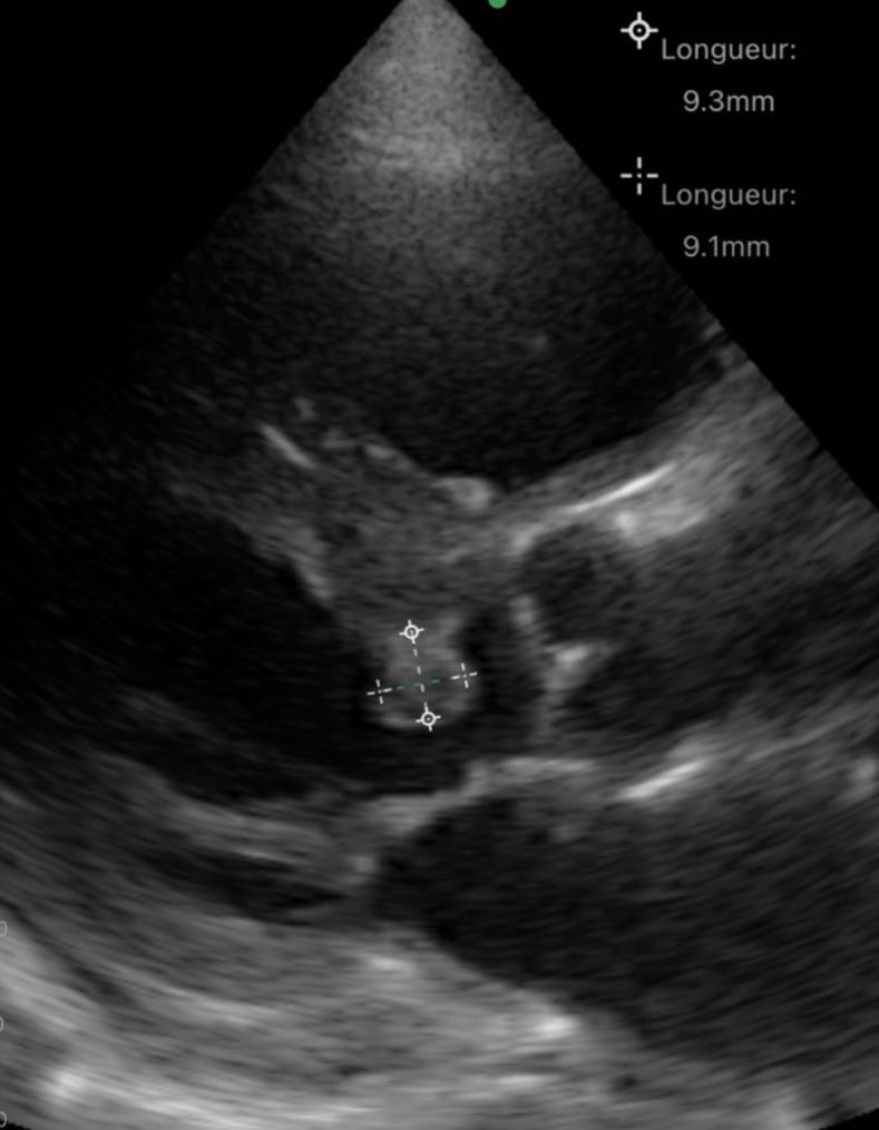
During the examination, the cardiologist identified a highly mobile intracardiac mass measuring approximately 9 mm, located within the left ventricular outflow tract (LVOT) immediately beneath the aortic valve.
Review of previous echocardiographic studies demonstrated that the lesion had already been present three years earlier, when it measured approximately 4 mm, indicating slow progressive enlargement rather than rapid growth.
Importantly, both the clinical presentation and echocardiographic findings showed no evidence of infective endocarditis.
At this stage, the lesion could not be confidently classified based solely on routine echocardiographic interpretation.
Rather than accepting diagnostic uncertainty or relying on an isolated opinion, the cardiologist initiated a structured discussion with artificial intelligence, using the echocardiographic findings as the starting point for collaborative differential diagnosis.
This approach reflects an important evolution in modern medical practice.
Artificial intelligence was not expected to replace physician expertise.
Instead, it was invited to participate in systematic clinical reasoning—much like discussing a complex case with an experienced colleague who continually explains the logic behind every diagnostic hypothesis.
This distinction is fundamental.
The value of AI lies not in producing immediate answers but in strengthening clinical thinking.
The effectiveness of this collaboration, however, depended entirely on one prerequisite: the availability of high-quality echocardiographic images obtained with reliable ultrasound equipment.
Without adequate visualization of lesion morphology, mobility, attachment site, and surrounding cardiac anatomy, meaningful AI analysis would simply not have been possible.
Physician–AI Dialogue: Step-by-Step Differential Diagnosis
The cardiologist presented the echocardiographic findings to artificial intelligence in the same way complex clinical cases are discussed during multidisciplinary consultations.
Rather than requesting a definitive diagnosis, the physician asked AI to analyze the available information and formulate the most likely differential diagnoses.
The response demonstrated one of artificial intelligence’s greatest strengths in clinical medicine: transparent reasoning instead of unsupported conclusions.
AI immediately acknowledged an important limitation. It explained that a single still echocardiographic image was insufficient for establishing a definitive diagnosis, emphasizing that additional imaging projections, lesion attachment, mobility, morphology, and video recordings would significantly improve diagnostic confidence.
This cautious approach mirrored the principles of evidence-based medicine and illustrated why AI should function as a clinical assistant rather than an autonomous decision-maker.
Based on the initial echocardiographic image, artificial intelligence identified several important characteristics:
- a small intracardiac mass measuring approximately 9 mm;
- remarkable mobility;
- probable pedunculated morphology;
- persistence over at least three years;
- gradual interval growth;
- absence of clinical evidence suggesting infective endocarditis.
Instead of proposing only one diagnosis, AI generated a structured differential diagnosis.
The leading hypothesis was papillary fibroelastoma, the most common benign cardiac tumor involving valvular and endocardial structures. Its small size, high mobility, slow growth, and pedunculated appearance were all compatible with this diagnosis.
However, artificial intelligence also considered several alternative possibilities.
These included Lambl’s excrescence, organized thrombus, cardiac myxoma, degenerative calcified lesions, and infective vegetation.
Importantly, each diagnostic possibility was accompanied by a detailed explanation rather than simply appearing as a list.
Artificial intelligence discussed why individual imaging features either supported or argued against every diagnosis.
This structured reasoning transformed the consultation from a simple answer into an educational discussion that could help physicians better understand the diagnostic process itself.
New Clinical Information Changes the Diagnostic Perspective
The physician then contributed additional echocardiographic observations obtained during the examination.
The lesion was attached to the muscular portion of the subaortic interventricular septum, approximately 10 mm below the aortic annulus.
A small pedicle could be identified.
The internal echogenicity appeared relatively homogeneous.
These new findings immediately influenced the diagnostic reasoning.
Artificial intelligence reassessed the entire differential diagnosis using the updated anatomical information.
Although papillary fibroelastoma remained the leading diagnosis, AI recognized that attachment to the muscular septum represented an atypical location compared with the more common valvular origin.
Rather than ignoring this discrepancy, artificial intelligence openly discussed it.
The lesion’s pronounced mobility, well-defined margins, homogeneous structure, pedunculated morphology, gradual enlargement over three years, and absence of infectious findings all continued to support papillary fibroelastoma.
Nevertheless, AI explained that an atypical left ventricular outflow tract myxoma should also remain under consideration because myxomas occasionally arise from the basal interventricular septum.
At the same time, organized thrombus became considerably less likely due to the lesion’s prolonged persistence and slow enlargement, while infective vegetation remained highly improbable in the complete absence of clinical evidence for endocarditis.
Following integration of the new information, artificial intelligence ranked the diagnostic possibilities as follows:
- Non-valvular papillary fibroelastoma.
- Atypical left ventricular outflow tract myxoma.
- Organized thrombus.
- Another rare benign intracardiac tumor.
Finally, AI suggested additional investigations that could further clarify the diagnosis if clinically appropriate, including transesophageal echocardiography, three-dimensional echocardiography, and cardiac magnetic resonance imaging.
This progression clearly illustrates why physician – AI collaboration is valuable.
Artificial intelligence did not simply provide a static answer.
Instead, it continuously refined its conclusions as new clinical information became available, closely resembling the dynamic reasoning process that occurs during discussions between experienced medical specialists.
At the same time, every refinement depended entirely on the physician’s ability to obtain precise echocardiographic information.
Without accurate visualization of lesion attachment, morphology, mobility, and surrounding anatomy using the SANTÉ INTÉGRALE wireless phased-array ultrasound probe, neither the physician nor artificial intelligence could have reached such a well-supported differential diagnosis.
This is perhaps the most important lesson of the entire case.
Artificial intelligence contributes analytical power.
The physician contributes clinical expertise.
High-quality ultrasound technology supplied by SANTÉ INTÉGRALE provides the diagnostic information that makes this collaboration possible.
Why High-Quality Ultrasound Technology Is Essential for AI-Assisted Diagnostics
This clinical case highlights an important principle of modern medical imaging: artificial intelligence is only as reliable as the diagnostic data it receives. AI cannot compensate for incomplete image acquisition or inadequate visualization. Every meaningful conclusion begins with a well-performed ultrasound examination.
In this case, the high-quality images obtained using the SANTÉ INTÉGRALE wireless phased-array Wi-Fi ultrasound probe enabled accurate assessment of the lesion’s mobility, attachment site, morphology, echogenicity, and interval growth. These imaging characteristics became the foundation for a structured AI-assisted differential diagnosis.
This illustrates why advanced ultrasound equipment remains indispensable in contemporary cardiology. Whether physicians choose a wireless ultrasound probe, a handheld ultrasound device, a portable ultrasound scanner, or a premium stationary ultrasound system, image quality directly influences diagnostic confidence and the usefulness of artificial intelligence.
SANTÉ INTÉGRALE Ultrasound Solutions for Modern Clinical Practice
Modern healthcare increasingly relies on flexible ultrasound solutions that combine excellent imaging performance with clinical mobility.
SANTÉ INTÉGRALE provides a comprehensive portfolio that includes wireless Wi-Fi ultrasound probes, handheld ultrasound devices, portable ultrasound scanners, phased-array cardiac probes, premium stationary ultrasound systems, and specialty transducers for multiple medical disciplines.
These systems support cardiology, emergency medicine, internal medicine, intensive care, vascular imaging, family medicine, obstetrics and gynecology, musculoskeletal ultrasound, urology, and many other specialties.
Portable ultrasound devices allow physicians to perform high-quality examinations at the bedside, in outpatient clinics, emergency departments, and remote healthcare settings, while premium stationary ultrasound systems remain the preferred choice for comprehensive echocardiographic assessment and advanced quantitative analysis.
Rather than competing, these technologies complement one another, allowing clinicians to select the most appropriate ultrasound solution for each clinical scenario while maintaining consistent diagnostic quality.
Conclusion
This case demonstrates that the future of cardiovascular imaging is based on collaboration between physician expertise, advanced ultrasound technology, and artificial intelligence.
The cardiologist acquired high-quality echocardiographic images using a SANTÉ INTÉGRALE wireless phased-array ultrasound probe, interpreted the findings critically, and engaged artificial intelligence in a structured clinical dialogue that evolved as additional anatomical information became available.
Artificial intelligence did not replace the physician. Instead, it strengthened clinical reasoning by organizing differential diagnoses, explaining diagnostic probabilities, and identifying the additional information needed for greater certainty.
For physicians, this case carries two practical messages. First, responsible AI can become a valuable clinical assistant when used critically and interactively. Second, reliable AI-assisted interpretation begins with high-quality ultrasound imaging. This is precisely where SANTÉ INTÉGRALE wireless ultrasound probes, portable ultrasound scanners, handheld ultrasound devices, and premium stationary ultrasound systems provide the technological foundation for confident, evidence-based clinical decision-making.
Frequently Asked Questions
Can a wireless ultrasound probe provide image quality suitable for advanced echocardiography?
Yes. Modern SANTÉ INTÉGRALE wireless phased-array ultrasound probes deliver high-quality cardiac imaging suitable for focused echocardiography, point-of-care ultrasound, bedside examinations, and many routine cardiovascular applications.
Can artificial intelligence diagnose cardiac tumors independently?
No. AI supports physicians by organizing differential diagnoses, identifying missing clinical information, and recommending additional investigations. Final diagnosis and patient management always remain the physician’s responsibility.
Why is physician – AI collaboration becoming increasingly important?
Artificial intelligence enhances clinical reasoning but depends on accurate imaging and clinical context. The combination of physician expertise, high-quality ultrasound technology, and structured AI analysis can improve diagnostic confidence in complex cases.
Why choose SANTÉ INTÉGRALE ultrasound systems?
SANTÉ INTÉGRALE offers a complete ecosystem of wireless ultrasound probes, handheld ultrasound devices, portable ultrasound scanners, and premium stationary ultrasound systems that combine mobility, excellent image quality, and clinical versatility for modern ultrasound practice.