
Meta Description:
Can a portable ultrasound scanner identify severe tricuspid regurgitation and right-sided heart failure? This clinical case demonstrates how a wireless ultrasound transducer from SANTÉ INTÉGRALE enabled rapid bedside cardiac assessment and detection of clinically significant valvular disease.
Suggested Image Alt Text:
Portable Wi-Fi ultrasound scanner showing severe tricuspid regurgitation on Doppler echocardiography in an elderly patient with atrial fibrillation.
Can a Portable Ultrasound Scanner Detect Severe Valvular Heart Disease?
Yes. Modern portable ultrasound scanners equipped with color Doppler and spectral Doppler capabilities can identify complex hemodynamic abnormalities, including severe tricuspid regurgitation, right atrial enlargement, venous congestion, and signs of right-sided heart failure.
This clinical case demonstrates how a wireless ultrasound transducer from SANTÉ INTÉGRALE was used to evaluate an elderly patient presenting with progressive weakness and lower-extremity edema. The examination revealed severe tricuspid valve insufficiency caused by right atrial dilatation associated with long-standing atrial fibrillation.
The case also highlights the growing role of portable ultrasound systems, handheld ultrasound devices, wireless ultrasound scanners, and smartphone-based ultrasound solutions in modern cardiovascular medicine. Technologies that once required access to a full-sized echocardiography laboratory are now increasingly available at the point of care, allowing clinicians to make informed decisions faster and more efficiently.
For healthcare professionals interested in expanding bedside imaging capabilities, it is useful to learn more about portable ultrasound systems and explore wireless ultrasound solutions currently used in contemporary clinical practice.
Clinical Case
An 83-year-old male patient presented with complaints of severe generalized weakness and progressive swelling of both lower limbs.
According to the patient, symptoms had gradually worsened over several months. He reported reduced exercise tolerance, increasing fatigue during routine daily activities, intermittent palpitations, and shortness of breath during moderate exertion. Lower-extremity edema became more pronounced in the evenings and progressively limited mobility.
The patient’s medical history included:
- Long-standing atrial fibrillation
- Arterial hypertension
- Chronic heart failure
- Advanced age with multiple cardiovascular risk factors
Physical examination revealed bilateral peripheral edema and clinical signs suggestive of systemic venous congestion. Given the patient’s symptoms and cardiovascular history, rapid assessment of cardiac structure and hemodynamics was considered essential.
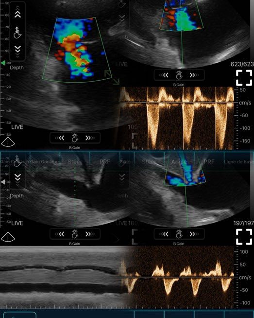
A bedside echocardiographic examination was performed using a portable ultrasound scanner from SANTÉ INTÉGRALE.
For cardiac imaging, the Phased Array Wi-Fi Mini Ultrasound Scanner Premium with HPRF and CW represents an ideal solution. However, the examination could also be performed using the Universal Premium 3-in-1 Wi-Fi Mini Ultrasound Scanner (192 Elements) or the 3-in-1 Wireless Ultrasound Probe combining Linear, Convex, and Phased Array technologies, both of which provide exceptional versatility for physicians working across multiple clinical specialties.
Ultrasound Examination Methodology
The examination was conducted using:
- B-mode imaging
- Color Doppler imaging
- Pulsed-wave Doppler
- Continuous-wave Doppler
These modalities allowed comprehensive assessment of:
- Right atrial dimensions
- Right ventricular morphology
- Tricuspid valve structure and function
- Inferior vena cava (IVC)
- Hepatic venous flow
- Cardiac hemodynamics
- Signs of systemic venous congestion
The ability of a portable ultrasound scanner to provide real-time hemodynamic information is one of the major reasons why handheld ultrasound technology has become increasingly valuable in cardiology, emergency medicine, and internal medicine.
Ultrasound Findings
During examination using a SANTÉ INTÉGRALE mini ultrasound scanner, the cause of the patient’s symptoms was identified as severe tricuspid valve insufficiency resulting from right atrial dilatation associated with atrial fibrillation.
Key echocardiographic signs of severe tricuspid regurgitation included:
- A large PISA zone
- Laminar spectral Doppler signal
- Hepatic vein flow reversal
- Dilatation of the inferior vena cava with absent inspiratory collapse
Color Doppler imaging demonstrated a broad regurgitant jet extending from the tricuspid valve into the right atrium.
The regurgitant flow occupied a substantial portion of the right atrial cavity, indicating advanced valvular insufficiency. Spectral Doppler analysis revealed a dense and laminar regurgitant signal consistent with severe tricuspid regurgitation.
One of the most important findings was the presence of a prominent proximal isovelocity surface area (PISA). PISA analysis remains a well-established echocardiographic parameter for assessing the severity of valvular regurgitation. A large PISA zone generally reflects a significant regurgitant orifice and increased regurgitant volume.
These findings strongly suggested advanced functional tricuspid regurgitation with substantial hemodynamic consequences.
Assessment of Hepatic Veins and Inferior Vena Cava
Evaluation of hepatic venous flow provided additional evidence supporting the diagnosis.
Under normal physiological conditions, blood flows continuously toward the right atrium. In severe tricuspid regurgitation, however, part of the blood volume is pushed backward into the venous circulation during systole.
This patient demonstrated clear hepatic vein flow reversal, one of the most specific echocardiographic indicators of severe tricuspid regurgitation.
The inferior vena cava was markedly dilated and showed virtually no collapse during inspiration.
In healthy individuals, inspiratory collapse of the inferior vena cava reflects normal right atrial pressure. Lack of collapse is generally associated with elevated right-sided filling pressures and systemic venous congestion.
Together, these findings confirmed advanced right-sided hemodynamic overload.
Pathophysiological Mechanism
Atrial fibrillation is often viewed primarily as a rhythm disorder. However, long-term atrial fibrillation also produces structural remodeling of cardiac chambers.
In this patient, chronic atrial fibrillation resulted in progressive enlargement of the right atrium. As the right atrium expanded, the tricuspid annulus became dilated.
This annular enlargement prevented complete leaflet coaptation during systole, leading to functional tricuspid regurgitation.
The resulting pathophysiological cascade can be summarized as follows:
Atrial fibrillation → Right atrial dilatation → Tricuspid annular dilatation → Severe tricuspid regurgitation → Elevated venous pressure → Systemic venous congestion → Peripheral edema and weakness → Progressive heart failure
This mechanism explains the patient’s symptoms remarkably well and illustrates why advanced echocardiographic assessment remains critical in elderly patients with unexplained edema and functional decline.
Why This Clinical Case Matters
Severe tricuspid regurgitation has historically received less attention than left-sided valvular diseases. However, growing evidence demonstrates that significant tricuspid valve insufficiency is associated with:
- Increased hospitalization rates
- Reduced quality of life
- Progressive right heart failure
- Higher mortality risk
Early recognition can significantly influence patient management and therapeutic decision-making.
This case demonstrates how a portable ultrasound scanner can rapidly identify clinically significant valvular disease at the bedside without requiring immediate access to a traditional echocardiography laboratory.
For clinicians evaluating point-of-care imaging solutions, cases like this emphasize the practical value of wireless ultrasound technology in everyday patient care.
The Role of Portable Ultrasound Systems in Modern Cardiology
Portable ultrasound technology has evolved considerably over the past decade.
Modern handheld ultrasound devices now offer:
- High-resolution imaging
- Color Doppler capabilities
- Spectral Doppler analysis
- Wireless connectivity
- Smartphone and tablet integration
- Rapid bedside deployment
As a result, a portable ultrasound scanner is increasingly considered an extension of the physical examination.
The ability to perform ultrasound directly at the point of care can improve diagnostic efficiency, reduce delays, and support more informed clinical decisions.
Healthcare professionals may wish to explore wireless ultrasound scanners, check available ultrasound devices, or see our ultrasound equipment catalog when evaluating solutions for cardiac imaging and bedside diagnostics.
Why the Universal 3-in-1 Wireless Ultrasound Probe Is Particularly Relevant
Although phased-array probes remain the preferred option for dedicated echocardiography, the Universal Premium 3-in-1 Wi-Fi Mini Ultrasound Scanner (192 Elements) offers unique advantages.
By combining phased-array, convex, and linear imaging capabilities within a single device, it enables clinicians to perform:
- Cardiac ultrasound
- Vascular ultrasound
- Abdominal ultrasound
- Musculoskeletal ultrasound
- Point-of-care examinations
- Emergency assessments
This versatility is especially valuable for physicians working in emergency departments, outpatient clinics, rural healthcare settings, and multidisciplinary practices.
The device represents a practical example of how modern wireless ultrasound scanners can support multiple clinical workflows while minimizing equipment requirements.
SANTÉ INTÉGRALE and Portable Ultrasound Solutions
SANTÉ INTÉGRALE is a provider of portable ultrasound solutions for healthcare professionals.
Its portfolio includes wireless ultrasound transducers, handheld ultrasound devices, portable ultrasound systems, and advanced point-of-care imaging technologies used across numerous specialties, including:
- Cardiology
- Emergency medicine
- Internal medicine
- Family medicine
- Gastroenterology
- Urology
- Endocrinology
- Musculoskeletal medicine
These technologies are increasingly adopted by physicians, clinics, distributors, and healthcare organizations seeking flexible and efficient diagnostic imaging solutions.
As portable imaging continues to evolve, wireless ultrasound technology is becoming an essential component of modern clinical practice.
Conclusion
This clinical case demonstrates the diagnostic value of portable echocardiography in an elderly patient with atrial fibrillation, severe tricuspid regurgitation, and symptoms of right-sided heart failure.
Using a portable ultrasound scanner from SANTÉ INTÉGRALE, clinicians were able to identify key echocardiographic findings including a large PISA zone, significant regurgitant flow, hepatic vein reflux, right atrial enlargement, and a dilated inferior vena cava without respiratory collapse.
The case highlights how wireless ultrasound technology can facilitate rapid bedside diagnosis and improve clinical decision-making.
Whether using the Phased Array Wi-Fi Mini Ultrasound Scanner Premium with HPRF and CW or the Universal Premium 3-in-1 Wi-Fi Mini Ultrasound Scanner, modern portable ultrasound systems provide clinicians with powerful diagnostic capabilities that were once available only on traditional cart-based equipment.
Frequently Asked Questions
In an elderly patient with atrial fibrillation and unexplained peripheral edema, what is the most practical first step in bedside ultrasound assessment?
In real-world practice, the priority is not immediately focusing on the valve, but identifying systemic venous congestion patterns. The first and most informative step is assessment of the inferior vena cava, right atrium size, and hepatic venous flow. A portable Wi-Fi ultrasound scanner such as SANTÉ INTÉGRALE allows rapid bedside confirmation of right-sided volume overload, often before full echocardiography is available. This early stratification can significantly accelerate clinical decision-making.
How does a portable Wi-Fi ultrasound scanner change clinical confidence in diagnosing severe tricuspid regurgitation?
Clinical confidence increases when multiple Doppler-based markers are obtained at the bedside in real time. In severe tricuspid regurgitation, findings such as a large PISA zone, systolic hepatic vein flow reversal, and a non-collapsing IVC often align into a consistent hemodynamic picture. A modern wireless ultrasound probe enables the clinician to integrate these findings immediately, reducing diagnostic delay and supporting early therapeutic decisions even before formal echocardiography.
Why is hepatic vein flow reversal considered more diagnostically powerful than visualizing the regurgitant jet alone?
Because the regurgitant jet describes the valvular lesion, while hepatic vein flow reversal reflects the systemic consequence of right atrial pressure overload. In clinical practice, this distinction is critical. Hepatic vein reversal indicates that tricuspid regurgitation is not merely anatomical but hemodynamically significant, with transmission of pressure into the venous system — a key marker of advanced disease.
Can a wireless ultrasound probe integrated with a smartphone provide sufficient information to guide early management decisions?
Yes, in appropriately selected cases. While it does not replace a comprehensive echocardiography lab study, it can provide actionable bedside information. In patients with suspected right-sided heart failure, a smartphone-connected Wi-Fi ultrasound probe can immediately differentiate cardiac venous congestion from non-cardiac causes of edema, allowing earlier initiation of diuretic or hemodynamic management strategies.
In atrial fibrillation, why does tricuspid regurgitation often remain underdiagnosed until advanced stages?
Because the disease evolves gradually through right atrial remodeling and annular dilation, symptoms remain nonspecific for a long period. Patients often present only when systemic congestion becomes clinically obvious. This delayed presentation is exactly where portable ultrasound becomes valuable — it allows detection of structural and functional changes at the point of care, before irreversible decompensation develops.
What additional clinical value does a 3-in-1 wireless ultrasound probe provide beyond focused cardiac imaging?
A 3-in-1 probe (linear, convex, and phased array) enables the clinician to immediately expand the diagnostic scope beyond the heart. In patients with severe tricuspid regurgitation, this allows simultaneous assessment of:
- venous congestion in the abdomen,
- hepatic vein dilation,
- pleural effusion,
- peripheral venous status.
This transforms a single-organ assessment into a whole-body congestion evaluation, improving diagnostic completeness in bedside practice.
When does portable ultrasound become essential rather than optional in suspected right heart failure?
It becomes essential when the clinical question is no longer “Is there heart disease?” but rather “Is this patient currently decompensated?”. In such cases, delays associated with referral-based imaging can affect management decisions. A portable Wi-Fi ultrasound scanner from SANTÉ INTÉGRALE enables immediate confirmation of congestion status, helping prioritize urgent treatment pathways.
Why are clinicians increasingly adopting wireless ultrasound systems instead of relying solely on cart-based echocardiography?
Because modern clinical workflows require speed, accessibility, and repeatability. Wireless ultrasound systems allow repeated bedside assessments without logistical constraints. This is particularly important in elderly patients, emergency settings, and outpatient follow-up, where hemodynamic status can change rapidly. As a result, portable ultrasound is increasingly functioning as an extension of the physical examination rather than a separate diagnostic event.