
Can a handheld ultrasound scanner identify severe structural heart disease in patients with malignant hypertension? Increasingly, the answer is yes. Modern handheld ultrasound systems have transformed cardiovascular imaging by bringing echocardiography directly to the bedside. Today, point-of-care ultrasound (POCUS) allows clinicians to evaluate left ventricular hypertrophy, ventricular dilation, regional wall motion abnormalities, and systolic dysfunction within minutes, helping accelerate clinical decision-making and improve patient management.
For cardiologists, internal medicine physicians, emergency care specialists, healthcare managers, and medical facilities, portable ultrasound has become more than a convenient imaging solution. It is an increasingly valuable diagnostic tool capable of providing clinically meaningful information during the initial patient encounter.
This clinical case demonstrates how point-of-care echocardiography performed with a wireless handheld ultrasound scanner contributed to the detection of advanced ischemic heart disease in a patient with malignant hypertension and progressive symptoms of heart failure.
Understanding Malignant Hypertension and Its Impact on the Heart
Malignant hypertension is one of the most severe forms of arterial hypertension and is associated with progressive damage to multiple organs. Persistent elevation of blood pressure places significant stress on the cardiovascular system, forcing the heart to adapt to increased afterload over many years.
Initially, the myocardium responds through compensatory thickening of the ventricular walls. This process, known as left ventricular hypertrophy (LVH), helps maintain cardiac output despite elevated systemic vascular resistance. However, prolonged hypertrophy eventually becomes maladaptive and contributes to adverse cardiac remodeling.
Potential consequences include:
- Left ventricular hypertrophy
- Left ventricular dilation
- Diastolic dysfunction
- Systolic dysfunction
- Reduced cardiac output
- Myocardial ischemia
- Chronic heart failure
- Increased cardiovascular mortality
Because symptoms may develop gradually, imaging plays a central role in identifying structural and functional cardiac abnormalities before irreversible deterioration occurs.
Clinical Case
A 62-year-old male with a long history of malignant arterial hypertension sought medical evaluation because of worsening shortness of breath during minimal physical activity and increasing fatigue. Although these symptoms are common in patients with advanced hypertensive heart disease, they may also indicate underlying ischemic myocardial damage.
To obtain immediate diagnostic information, clinicians performed point-of-care echocardiography using a wireless handheld ultrasound scanner.
The examination identified several important abnormalities:
Echocardiographic Parameter
| Finding
|
Interventricular septum
| 17–18 mm
|
Left ventricular end-diastolic diameter
| 59 mm
|
Lateral wall motion
| Akinesia
|
Other myocardial segments
| Diffuse hypokinesia
|
Velocity Time Integral (VTI)
| 14 cm
|
Cardiac output
| Reduced
|
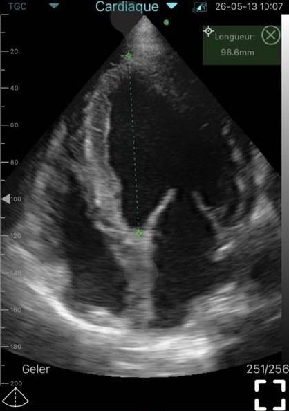
The findings demonstrated severe non-obstructive left ventricular hypertrophy, moderate ventricular dilation, significant regional wall motion abnormalities, and evidence of reduced pumping function.
Importantly, the combination of akinesia and diffuse hypokinesia suggested that the patient’s condition extended beyond hypertensive remodeling alone and raised suspicion of significant coronary artery disease.
Subsequent coronary angiography confirmed complete occlusion of the circumflex artery responsible for lateral wall infarction and critical 90% stenosis of the left anterior descending artery. Coronary stenting was successfully performed, and the patient entered long-term cardiology follow-up.
Why Point-of-Care Echocardiography Matters
The presented case illustrates one of the greatest advantages of point-of-care echocardiography: immediate access to clinically actionable information.
Without portable ultrasound, this patient’s symptoms could initially have been attributed solely to chronic hypertension and heart failure. Instead, bedside imaging revealed findings suggestive of ischemic myocardial injury, prompting additional investigation and ultimately leading to life-saving intervention.
Point-of-care echocardiography enabled clinicians to identify:
- Severe myocardial hypertrophy
- Ventricular enlargement
- Regional wall motion abnormalities
- Reduced cardiac performance
- Signs consistent with ischemic heart disease
These findings substantially changed the diagnostic pathway and accelerated referral for coronary angiography.
Echocardiographic Analysis of the Cardiac Findings Severe Left Ventricular Hypertrophy
Left ventricular hypertrophy remains one of the most important manifestations of chronic hypertension.
In this patient, septal thickness reached 17–18 mm, indicating advanced myocardial remodeling. Such hypertrophy develops because the myocardium must generate greater force to overcome persistently elevated blood pressure.
Clinical implications of severe LVH include:
- Increased myocardial oxygen demand
- Reduced coronary perfusion reserve
- Elevated risk of arrhythmias
- Greater likelihood of heart failure
- Higher incidence of cardiovascular events
Portable echocardiography allows clinicians to detect these structural changes rapidly during routine patient evaluation.
Ventricular Dilation
The observed left ventricular end-diastolic diameter of 59 mm demonstrated moderate chamber enlargement.
When hypertrophy and dilation occur simultaneously, the myocardium is often transitioning from compensation toward progressive dysfunction. This stage of remodeling may be associated with worsening exercise tolerance, reduced cardiac efficiency, and declining prognosis.
Regional Wall Motion Abnormalities
Among all findings, lateral wall akinesia carried particularly important clinical significance.
Regional wall motion abnormalities frequently indicate:
- Previous myocardial infarction
- Ongoing ischemia
- Coronary artery obstruction
- Myocardial scar formation
The detection of akinesia using handheld ultrasound directly contributed to the decision to perform coronary angiography.
Reduced Cardiac Output
The measured VTI of 14 cm suggested diminished stroke volume and impaired cardiac output.
This functional abnormality correlated with the patient’s symptoms of fatigue and exertional dyspnea, demonstrating how portable ultrasound can connect structural findings with physiological consequences.
Handheld Ultrasound as an Extension of the Physical Examination
Traditional cardiovascular examination remains essential but has limitations when evaluating structural heart disease.
Modern handheld ultrasound increasingly functions as an extension of bedside assessment by allowing physicians to visualize cardiac anatomy and function during the consultation itself.
Advantages include:
Rapid Assessment
Clinicians receive immediate diagnostic information without waiting for a separate imaging appointment.
Improved Clinical Decision-Making
Real-time imaging supports more informed referral and treatment decisions.
Repeatability
Portable echocardiography can be repeated frequently to monitor disease progression and treatment response.
Greater Accessibility
Handheld ultrasound systems expand access to cardiac imaging in outpatient clinics, emergency settings, intensive care units, and mobile healthcare services.
Healthcare professionals interested in improving bedside diagnostics may benefit from learning more about portable ultrasound systems and integrating point-of-care ultrasound into everyday practice.
Applications of Portable Ultrasound in Modern Cardiology
The role of handheld ultrasound continues to expand throughout cardiovascular medicine.
Hypertensive Heart Disease
Portable echocardiography helps monitor:
- Myocardial thickness
- Chamber dimensions
- Ventricular function
- Cardiac remodeling
Heart Failure Evaluation
Bedside ultrasound supports assessment of:
- Ventricular performance
- Cardiac output
- Structural abnormalities
- Disease progression
Ischemic Heart Disease
Although coronary anatomy cannot be visualized directly, regional wall motion abnormalities may indicate significant coronary pathology requiring further investigation.
Post-Revascularization Monitoring
Patients who undergo coronary intervention often benefit from serial evaluation of ventricular function and remodeling.
Ambulatory Cardiology
Portable ultrasound allows cardiologists to combine clinical consultation and imaging within a single patient encounter.
Wireless Ultrasound Technology and Diagnostic Performance
Historically, many clinicians questioned whether handheld ultrasound could deliver image quality sufficient for reliable cardiac assessment.
Advances in transducer technology, wireless connectivity, signal processing, and software optimization have dramatically improved performance.
Modern wireless ultrasound scanners offer:
Feature
| Clinical Benefit
|
Wireless operation
| Improved mobility
|
High-resolution imaging
| Better visualization
|
Real-time assessment
| Faster diagnosis
|
Compact design
| Point-of-care usability
|
Multiple scanning applications
| Greater versatility
|
These improvements have significantly increased the value of handheld ultrasound in clinical cardiology.
Pros and Cons of Handheld Ultrasound in Cardiac Practice
Advantages
- Immediate bedside imaging
- Faster clinical decisions
- Improved workflow efficiency
- Enhanced patient access to diagnostics
- Repeat examinations without complex scheduling
- Useful for screening and follow-up
- Supports mobile and community healthcare
Limitations
- Does not replace comprehensive expert echocardiography
- Operator training remains essential
- Advanced quantitative measurements may require full-featured systems
- Some complex cardiac conditions still require referral imaging
For most clinicians, handheld ultrasound should be viewed as a complementary diagnostic tool that enhances rather than replaces traditional echocardiography.
Sante Integrale Handheld Ultrasound Solutions
Sante Integrale is a provider of wireless handheld ultrasound systems designed for modern clinical practice. These portable imaging solutions are used across cardiology, emergency medicine, critical care, family medicine, vascular diagnostics, obstetrics and gynecology, and point-of-care ultrasound applications.
The company’s portfolio includes wireless ultrasound scanners and multifunctional devices capable of supporting phased-array, convex, and linear imaging within a single portable platform.
Clinics and healthcare organizations seeking to modernize bedside imaging workflows may wish to explore wireless ultrasound solutions, learn more about portable ultrasound systems, see our ultrasound equipment catalog, or check available ultrasound devices suitable for point-of-care applications.
As a trusted partner in mobile ultrasound technology, Sante Integrale supports healthcare professionals who seek greater flexibility and accessibility in diagnostic imaging.
Conclusion
This clinical case highlights the important role of handheld ultrasound in the evaluation of patients with malignant hypertension and suspected cardiovascular complications.
Point-of-care echocardiography identified severe left ventricular hypertrophy, ventricular dilation, regional wall motion abnormalities, and reduced cardiac output. These findings led directly to coronary angiography, which confirmed advanced coronary artery disease and enabled timely intervention.
The growing adoption of portable ultrasound reflects a broader shift toward faster, more accessible, and more efficient diagnostic pathways. For cardiologists, clinics, healthcare managers, distributors, and medical buyers, handheld ultrasound offers a practical solution that combines mobility, diagnostic value, and clinical efficiency.
point-of-care ultrasound continues to evolve, portable echocardiography is expected to play an increasingly important role in the detection, monitoring, and management of cardiovascular disease.
Frequently Asked Questions
Can handheld ultrasound detect left ventricular hypertrophy?
Yes. Handheld ultrasound can accurately visualize myocardial thickening, chamber geometry, and structural changes associated with left ventricular hypertrophy.
Is point-of-care echocardiography useful for identifying myocardial ischemia?
Yes. Point-of-care echocardiography can detect regional wall motion abnormalities that may indicate myocardial ischemia or previous myocardial infarction.
Can portable ultrasound identify systolic dysfunction?
Yes. Portable ultrasound can assess ventricular contractility, stroke volume indicators, and overall systolic performance during bedside examination.
Who benefits from wireless ultrasound scanners in cardiology?
Cardiologists, emergency physicians, intensivists, internal medicine specialists, family physicians, healthcare managers, clinics, and medical buyers all benefit from portable cardiac imaging solutions.
Why are handheld ultrasound devices becoming more common?
Handheld ultrasound devices improve accessibility, reduce diagnostic delays, support point-of-care ultrasound workflows, and facilitate repeated examinations.
Can handheld ultrasound be used for echocardiography?
Yes. Modern handheld ultrasound systems can be effectively used for comprehensive echocardiographic assessment in many clinical settings. They enable evaluation of cardiac anatomy, ventricular function, myocardial hypertrophy, chamber dimensions, valvular abnormalities, and regional wall motion changes. Depending on the clinical scenario, handheld ultrasound may serve as a primary imaging tool or complement conventional ultrasound systems, while offering the advantages of portability, immediate bedside access, and rapid clinical decision-making.