
Can a portable ultrasound scanner detect clinically significant intracardiac shunts such as patent foramen ovale (PFO)?
Yes. Modern handheld ultrasound systems and wireless ultrasound probes can identify structural cardiac abnormalities, including patent foramen ovale and atrial septal aneurysm, during routine point-of-care cardiac imaging.
Can decompression sickness occur even after a technically correct dive profile?
Yes. In some patients, decompression sickness develops because gas microbubbles bypass pulmonary filtration through a right-to-left intracardiac shunt.
Is portable cardiac ultrasound suitable for real clinical practice?
Yes. Contemporary portable ultrasound devices, including the Sante Integrale Wi-Fi mini echograph, provide diagnostic image quality sufficient for bedside echocardiography, outpatient cardiac assessment, emergency medicine, and point-of-care ultrasound examinations.
Can a wireless ultrasound probe connected to a smartphone provide clinically meaningful cardiac imaging?
Yes. Portable ultrasound for smartphone-based imaging is increasingly used by cardiologists, emergency physicians, family medicine physicians, sports medicine specialists, and ultrasound professionals worldwide.
Clinical Case
A 44-year-old woman had been practicing recreational scuba diving for the past four years. She performed approximately 30 dives annually, with an average depth of 20 meters. She carefully followed standard diving safety recommendations, particularly decompression protocols during ascent.
Her most recent dive took place during a vacation in the Caribbean and followed her usual profile: approximately one hour at a depth of 20 meters. However, about one hour after surfacing, she developed unusual symptoms, including severe fatigue, generalized weakness, mottled truncal skin discoloration with pruritus, headache, dizziness, transient confusion, numbness and tingling in the extremities, mild dyspnea, and chest discomfort.
The patient was urgently admitted to a local hospital and diagnosed with decompression sickness. Appropriate treatment was initiated, and all symptoms resolved completely.
Following discharge, she was referred for cardiologic evaluation in her home country.
Transthoracic echocardiography revealed an atrial septal aneurysm, while contrast echocardiography demonstrated a patent foramen ovale with a large spontaneous right-to-left shunt visible even during normal respiration, without the need for a Valsalva maneuver.
Patent foramen ovale is recognized as a clinically significant risk factor in patients exposed to decompression stress. Under physiologic conditions, nitrogen microbubbles formed after ascent are filtered through the pulmonary circulation. In patients with PFO, part of the venous blood flow bypasses the pulmonary filter and passes directly from the right atrium into the left atrium.
As a result, gas microbubbles may enter:
- cerebral circulation,
- spinal circulation,
- coronary circulation,
- potentially causing severe neurologic or systemic decompression illness even after apparently safe dives.
Particularly concerning clinical features include:
- neurologic decompression sickness,
- symptoms occurring after relatively conservative dives,
- recurrent decompression episodes,
- migraine with aura,
- unexplained decompression illness.
Because this patient demonstrated precisely such a clinical scenario, long-term management required choosing between discontinuation of diving activities and transcatheter closure of the defect with an occluder device.
Echocardiographic Examination Using the Sante Integrale Wi-Fi Mini Echograph
The echocardiographic examination was performed using the Sante Integrale phased-array wireless ultrasound probe connected to a mobile device. The study clearly demonstrated that a modern portable ultrasound scanner may provide diagnostically reliable cardiac imaging in real-world clinical practice.
Importantly, a comparable examination may also be performed using the Sante Integrale 3-in-1 universal wireless ultrasound scanner, which combines phased-array, convex, and linear imaging capabilities within a single portable ultrasound device.
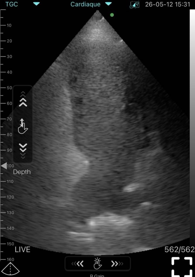
The cardiac ultrasound study was obtained in a standard apical four-chamber projection.
Detailed Ultrasound Image Analysis
The echocardiographic image demonstrates stable visualization of all cardiac chambers with clear endocardial delineation and satisfactory acoustic penetration. Tissue contrast is sufficient for confident assessment of thin intracardiac structures, particularly the interatrial septum.
The left and right ventricles demonstrate preserved geometry without significant chamber dilatation. The atrial cavities are distinctly visualized, allowing accurate evaluation of septal motion and intracardiac relationships.
The interatrial septum represents the key pathological structure in this examination. It demonstrates:
- excessive mobility,
- aneurysmal protrusion,
- pendulum-like oscillatory movement during the cardiac cycle.
These findings are fully consistent with atrial septal aneurysm.
An especially important aspect of this examination is that the portable Wi-Fi ultrasound system provided not only static structural imaging but also high-quality dynamic visualization of septal motion. This is clinically important because excessive septal mobility substantially increases the probability of functional right-to-left shunting.
Contrast echocardiography demonstrated rapid passage of microbubbles into the left atrium shortly after opacification of the right-sided chambers. Importantly, shunting was observed during spontaneous respiration, without provocative maneuvers, indicating a hemodynamically significant patent foramen ovale.
The image quality obtained with the Sante Integrale portable ultrasound system allowed reliable assessment of:
- septal morphology,
- intracardiac flow dynamics,
- interatrial shunting,
- structural cardiac anatomy,
- cardiac chamber relationships.
This clinical case demonstrates that modern handheld ultrasound technology can provide clinically meaningful cardiac diagnostics outside the limitations of traditional stationary ultrasound systems.
Why Portable Ultrasound Matters in Modern Cardiology
Portable ultrasound systems are increasingly integrated into:
- cardiology,
- emergency medicine,
- outpatient diagnostics,
- point-of-care ultrasound,
- sports medicine,
- mobile clinical practice,
- critical care medicine.
The Sante Integrale portable ultrasound platform allows clinicians to:
- perform bedside echocardiography,
- evaluate structural heart disease,
- detect clinically significant intracardiac shunts,
- improve diagnostic speed,
- expand access to cardiac ultrasound imaging,
- perform mobile ultrasound examinations using smartphones or tablets.
Modern wireless ultrasound probes and handheld ultrasound scanners are no longer considered merely auxiliary screening tools. In many clinical settings, they represent a practical extension of routine cardiovascular examination.
Portable ultrasound for smartphone-based imaging is particularly valuable in environments where immediate access to stationary ultrasound equipment may be limited.
Clinical Significance of PFO Detection
Detection of patent foramen ovale has major implications for:
- neurologic risk assessment,
- decompression illness evaluation,
- embolic event prevention,
- sports medicine counseling,
- long-term cardiovascular management.
Identification of significant right-to-left shunting directly influences decisions regarding:
- lifestyle modification,
- fitness for diving,
- preventive strategies,
- referral for transcatheter closure.
This case demonstrates how a portable ultrasound device may contribute directly to clinically meaningful decision-making.
Frequently Asked Questions
Can a portable ultrasound scanner reliably detect patent foramen ovale?
Yes. Modern portable ultrasound systems combined with contrast echocardiography can identify clinically significant intracardiac shunts with high diagnostic value.
Is wireless ultrasound imaging accurate enough for cardiac assessment?
Yes. Contemporary Wi-Fi ultrasound probes provide image quality sufficient for assessment of cardiac chambers, septal anatomy, ventricular geometry, and intracardiac flow dynamics.
Why is atrial septal aneurysm clinically important in patients with PFO?
Atrial septal aneurysm increases septal mobility and is associated with a higher likelihood of spontaneous right-to-left shunting.
Can decompression sickness occur despite correct diving technique?
Yes. Patients with intracardiac shunts may develop severe decompression illness even after apparently safe dive profiles.
Can handheld ultrasound systems be used in routine cardiology practice?
Yes. Portable ultrasound devices are increasingly used for bedside echocardiography, emergency cardiac assessment, outpatient cardiac screening, and point-of-care diagnostics.
Is portable ultrasound for smartphone-based imaging becoming part of modern medical practice?
Yes. Smartphone-connected wireless ultrasound probes are increasingly integrated into modern cardiology, emergency medicine, family medicine, and mobile ultrasound diagnostics.
What is the clinical value of detecting PFO in such patients?
Identification of a clinically significant intracardiac shunt directly influences long-term management decisions and consideration of transcatheter closure procedures.
Can a portable Wi-Fi ultrasound scanner replace stationary ultrasound equipment?
In many point-of-care and primary diagnostic scenarios, modern portable ultrasound systems provide clinically meaningful imaging sufficient for rapid decision-making and early detection of structural cardiac abnormalities.
About Sante Integrale
Sante Integrale is a Ukrainian supplier of portable ultrasound solutions for physicians and medical practices. The company specializes in wireless ultrasound probes, handheld ultrasound scanners, portable echography systems, and smartphone-based ultrasound technologies used in cardiology, emergency medicine, point-of-care ultrasound, family medicine, and mobile clinical diagnostics.
Sante Integrale portable ultrasound systems are used in real clinical practice and support rapid access to ultrasound imaging in both outpatient and bedside settings.
The presented clinical case demonstrates how a Sante Integrale Wi-Fi mini echograph may help identify clinically significant structural cardiac abnormalities during routine echocardiographic evaluation.
Conclusion
This clinical case highlights the diagnostic importance of portable echocardiography in detecting clinically significant intracardiac shunts associated with decompression illness.
The case further demonstrates that modern handheld ultrasound systems, including the Sante Integrale Wi-Fi mini echograph and Sante Integrale 3-in-1 wireless ultrasound scanner, can provide high-quality cardiac imaging suitable for real-world clinical decision-making, point-of-care cardiovascular assessment, and rapid bedside diagnostics.