
Can a portable ultrasound scanner identify serious structural heart disease during a routine outpatient consultation? Modern wireless ultrasound technology increasingly demonstrates that it can. In this clinical case, an ultra-portable Wi-Fi ultrasound scanner from Sante Integrale enabled immediate visualization of severe concentric left ventricular hypertrophy and raised strong suspicion of hypertrophic cardiomyopathy during the patient’s first evaluation.
Portable echocardiography is no longer limited to rapid screening alone. A modern portable ultrasound device connected to a smartphone can provide clinically meaningful cardiac imaging, support early diagnostic decisions, and accelerate referral for advanced cardiology assessment. In cardiology, emergency medicine, family medicine, and outpatient care, portable ultrasound systems are becoming an integral part of real-world clinical workflows.
This case also illustrates how a wireless ultrasound scanner can improve access to cardiac imaging outside traditional diagnostic laboratories. High-quality visualization, adequate penetration depth, and immediate bedside availability make portable ultrasound technology increasingly valuable for physicians managing patients with potentially serious cardiovascular disease.
Clinical Case
A 43-year-old woman presented with exertional dyspnea, episodic palpitations, and intermittent chest discomfort. According to the patient, symptoms had gradually progressed over several months. She also reported reduced exercise tolerance, occasional weakness after climbing stairs, and shortness of breath during brisk walking.
The patient had not undergone recent cardiology evaluation because she attributed her symptoms to stress and fatigue. However, the family history raised additional concern. Her father had arterial hypertension, and a close relative reportedly died suddenly at a relatively young age due to suspected cardiac disease.
During the initial examination, blood pressure was 145/90 mmHg and heart rate was 96 beats per minute. Cardiac auscultation revealed a moderate systolic murmur over the apex. Given the patient’s symptoms and family history, immediate cardiac ultrasound examination was performed using a portable ultrasound scanner equipped with a phased array Wi-Fi probe.
The examination was conducted directly during the consultation without referring the patient to a separate echocardiography laboratory. This approach demonstrates one of the key advantages of portable ultrasound systems in contemporary practice: the ability to integrate diagnostic imaging into real-time clinical decision-making.
The phased array ultrasound probe used in this case was optimized for cardiac imaging. The portable ultrasound device provided adequate penetration depth and sector scanning quality required for transthoracic echocardiography.
Clinicians who want to learn more about portable ultrasound systems increasingly prioritize devices that combine mobility, rapid deployment, and clinically reliable cardiac imaging.
Why Portable Echocardiography Is Becoming Essential in Modern Cardiology
Portable echocardiography has significantly changed the way clinicians approach cardiovascular assessment. In many outpatient and emergency settings, physicians need rapid structural evaluation of the heart before making further diagnostic or therapeutic decisions.
A wireless ultrasound scanner offers several important clinical advantages:
- immediate access to cardiac imaging;
- reduced delay between consultation and diagnosis;
- improved triage in symptomatic patients;
- integration of imaging into bedside examination;
- greater mobility for outpatient and emergency care;
- support for point-of-care ultrasound protocols.
Modern portable ultrasound systems are increasingly used in:
- cardiology clinics;
- emergency departments;
- outpatient medical centers;
- family medicine practices;
- intensive care units;
- mobile and field medicine.
Many healthcare providers now explore wireless ultrasound solutions to improve workflow efficiency while maintaining diagnostic value.
Ultrasound Examination Methodology Using a Wi-Fi Ultrasound Scanner
The cardiac ultrasound examination was performed using standard echocardiographic windows with an ultra-portable ultrasound scanner connected to a smartphone. The portable ultrasound device allowed immediate access to diagnostic imaging without the need for a conventional stationary ultrasound machine.
The wireless ultrasound probe provided clear visualization of:
- left ventricular geometry;
- interventricular septum thickness;
- myocardial structure;
- chamber dimensions;
- valve morphology;
- global wall contractility.
The examination included:
- B-mode imaging;
- sector scanning;
- basic transthoracic echocardiography;
- myocardial thickness measurements;
- evaluation of left ventricular geometry;
- assessment of intracardiac hemodynamics;
- visual analysis of valvular structures.
Particular attention was directed toward the interventricular septum, posterior wall thickness, and the relationship between myocardial thickness and ventricular cavity size. These parameters are essential when differentiating hypertrophic cardiomyopathy from hypertensive myocardial remodeling.
The quality of visualization achieved with the portable ultrasound scanner was clinically significant. Despite the compact format, the device provided sufficient image resolution and tissue contrast to support accurate bedside cardiac assessment.
Clinicians who want to learn more about portable ultrasound systems increasingly recognize the role of wireless imaging technologies in rapid cardiovascular evaluation. Many healthcare providers also explore portable cardiac ultrasound workflows to improve bedside diagnostics and streamline patient management.
Detailed Echocardiographic Image Analysis
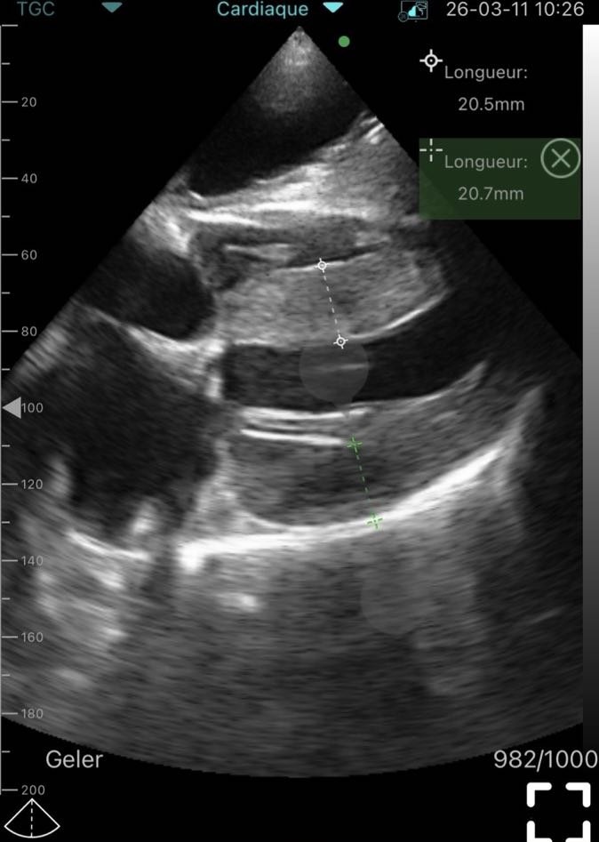
The echocardiographic images obtained with the portable ultrasound scanner demonstrated marked concentric thickening of the left ventricular myocardium.
Measured myocardial thickness exceeded 20 mm, representing severe pathological hypertrophy. The images also revealed a relatively reduced left ventricular cavity size combined with diffuse myocardial thickening.
Several important echocardiographic findings supported the diagnosis of hypertrophic cardiomyopathy:
- pronounced thickening of the interventricular septum;
- concentric hypertrophic remodeling;
- reduced left ventricular cavity dimensions;
- preserved visualization of valvular structures;
- adequate acoustic penetration and tissue contrast.
Endocardial borders remained clearly visible, allowing precise placement of measurement calipers even within the portable imaging format. Measurements obtained during the examination ranged from approximately 20.5 to 20.7 mm.
Another clinically important feature was the homogeneous echogenic appearance of the hypertrophied myocardium without evidence of focal scar defects or regional wall thinning.
The relatively narrow ventricular cavity observed during the examination is commonly associated with hypertrophic cardiomyopathy and may contribute to:
- diastolic dysfunction;
- impaired ventricular filling;
- progressive exertional dyspnea;
- reduced exercise tolerance.
The phased array Wi-Fi ultrasound probe also demonstrated stable visualization quality despite the technical challenges associated with cardiac imaging. For portable echocardiography, image quality remains one of the most critical factors determining diagnostic reliability.
The examination did not reveal major structural valvular abnormalities. However, indirect signs of diastolic overload suggested functional hemodynamic consequences of myocardial hypertrophy.
These findings justified urgent referral for:
- comprehensive expert-level echocardiography;
- ECG monitoring;
- arrhythmia risk assessment;
- advanced cardiology evaluation.
This clinical case demonstrates that a modern portable ultrasound scanner can function as a valuable primary imaging tool rather than merely a supplementary screening device.
Differential Diagnosis: Hypertrophic Cardiomyopathy vs Hypertensive Remodeling
An important part of image interpretation involved differentiating hypertrophic cardiomyopathy from secondary myocardial hypertrophy caused by arterial hypertension.
Several factors favored hypertrophic cardiomyopathy:
Clinical and Imaging Feature | Diagnostic Significance |
Myocardial thickness greater than 20 mm | Severe pathological hypertrophy |
Concentric remodeling pattern | Suggestive of structural myocardial disease |
Reduced left ventricular cavity | Common in hypertrophic cardiomyopathy |
Symptomatic presentation | Clinically significant disease |
Relatively young patient age | Less typical for isolated hypertensive remodeling |
Family history of cardiac disease | Possible hereditary cardiomyopathy |
Although arterial hypertension may contribute to myocardial remodeling, the degree of hypertrophy and the overall echocardiographic pattern strongly supported primary cardiomyopathy.
Portable ultrasound technology increasingly assists clinicians in identifying these abnormalities during initial patient assessment. Today, a wireless ultrasound scanner can provide high-quality cardiac ultrasound imaging without dependence on large stationary systems. This is especially important in outpatient cardiology, emergency medicine, and bedside cardiac assessment.
Clinical Importance of Early Ultrasound Diagnosis in Hypertrophic Cardiomyopathy
Hypertrophic cardiomyopathy is a primary myocardial disorder characterized by abnormal thickening of the cardiac muscle. The disease may remain undiagnosed for years, especially in patients with nonspecific symptoms.
Early ultrasound diagnosis is therefore critically important.
Portable echocardiography allows clinicians to evaluate cardiac structure during the first patient encounter, potentially accelerating diagnosis and specialist referral. In symptomatic patients, rapid access to cardiac imaging may substantially reduce delays in identifying potentially life-threatening disease.
In this case, the wireless ultrasound probe became an effective tool for early detection of significant structural pathology. Immediate echocardiographic assessment prompted rapid referral for specialized cardiology management.
Portable ultrasound systems are particularly valuable in clinical environments where immediate bedside cardiac assessment influences patient routing and early treatment decisions.
Portable ultrasound systems are particularly valuable in:
- outpatient cardiology;
- small private clinics;
- family medicine;
- emergency medicine;
- remote consultations;
- healthcare environments with limited imaging infrastructure.
Many clinicians now check available ultrasound devices specifically for bedside cardiac assessment capabilities before integrating them into routine practice.
Advantages of Portable Ultrasound Devices in Real Clinical Practice
The growing adoption of portable ultrasound scanners is driven not only by mobility but also by clinical practicality.
Key Advantages of Portable Ultrasound Systems
Advantage | Clinical Benefit |
Smartphone connectivity | Rapid deployment during consultation |
Wireless operation | Improved mobility and workflow |
Compact size | Easier use in outpatient and emergency settings |
Real-time visualization | Faster clinical decisions |
Cardiac imaging capability | Earlier detection of structural heart disease |
Point-of-care integration | Improved patient management efficiency |
Modern handheld ultrasound systems are increasingly integrated into point-of-care ultrasound protocols because they provide fast access to cardiovascular ultrasound diagnostics directly during patient examination.
Portable ultrasound technology can support:
- rapid triage;
- bedside diagnostics;
- follow-up examinations;
- screening of symptomatic patients;
- procedural guidance in selected settings.
Clinicians who explore wireless ultrasound scanners often prioritize devices capable of reliable cardiac visualization because cardiovascular assessment remains one of the most demanding areas of point-of-care imaging.
The Role of Sante Integrale in Portable Ultrasound Solutions
Sante Integrale is a trusted supplier of portable ultrasound equipment for healthcare professionals and medical institutions. The company provides modern wireless ultrasound solutions designed for use across multiple clinical specialties.
Portable ultrasound systems available from Sante Integrale are increasingly used in:
- cardiology;
- emergency medicine;
- outpatient diagnostics;
- private medical practice;
- mobile healthcare services.
The combination of mobility, high-quality visualization, and rapid smartphone connectivity makes modern portable ultrasound devices highly practical for daily clinical work.
Healthcare professionals seeking to see our ultrasound equipment catalog often focus on compact systems that combine portability with clinically meaningful imaging performance. Clinics that explore wireless ultrasound scanners increasingly value systems optimized for portable echocardiography and rapid bedside imaging.
Clinical Significance of This Case
This clinical case highlights several important aspects of modern portable echocardiography:
- the importance of early cardiac ultrasound screening;
- the diagnostic value of portable ultrasound scanners;
- the ability of wireless ultrasound probes to detect clinically significant structural disease;
- the usefulness of point-of-care echocardiography during the initial consultation;
- the role of portable imaging in accelerating patient referral pathways;
- the growing clinical importance of smartphone-based ultrasound systems.
The case also demonstrates that portable ultrasound technology is increasingly capable of providing actionable diagnostic information in cardiology.
Future Perspectives of Portable Cardiac Ultrasound
The role of portable ultrasound systems in cardiovascular medicine continues to expand. Improvements in image processing, wireless connectivity, and probe miniaturization are making handheld ultrasound increasingly accessible to physicians across multiple specialties.
Several trends are likely to shape the future of portable echocardiography:
- broader integration into primary care;
- increased use in emergency triage;
- expansion of telemedicine-supported imaging;
- greater adoption in rural healthcare settings;
- improved AI-assisted image interpretation;
- wider use in preventive cardiovascular screening.
As portable ultrasound devices continue to evolve, clinicians may increasingly rely on bedside imaging for earlier recognition of structural heart disease.
Healthcare organizations that learn more about portable ultrasound systems are also recognizing their potential role in improving diagnostic accessibility and workflow efficiency.
Conclusion
This clinical case demonstrates that a portable ultrasound scanner equipped with a phased array Wi-Fi probe can identify severe structural cardiac pathology during routine clinical evaluation.
The ultra-portable ultrasound system used in this examination enabled immediate visualization of marked concentric left ventricular hypertrophy consistent with hypertrophic cardiomyopathy. Rapid bedside echocardiography accelerated diagnostic decision-making and facilitated timely referral for specialized cardiology assessment.
Modern wireless ultrasound technology is increasingly transforming clinical practice by bringing high-quality imaging directly to the point of care. Portable echocardiography is becoming an essential component of contemporary cardiovascular assessment in outpatient medicine, emergency care, and mobile healthcare environments.
Clinicians who explore wireless ultrasound solutions increasingly recognize the importance of combining mobility with clinically reliable visualization quality.
Frequently Asked Questions
The following frequently asked questions are designed to address common clinical and technical considerations related to portable ultrasound scanners, portable echocardiography, and wireless cardiac imaging systems.
Can a portable ultrasound scanner reliably detect hypertrophic cardiomyopathy during outpatient evaluation?
Yes. Modern portable ultrasound scanners with phased array probes can visualize significant myocardial hypertrophy, ventricular geometry changes, and other structural abnormalities associated with hypertrophic cardiomyopathy.
Is portable echocardiography useful during outpatient consultations and point-of-care cardiac assessment?
Portable echocardiography is highly useful during outpatient visits because it allows immediate cardiac assessment without waiting for referral to a separate imaging department.
Why is a phased array Wi-Fi ultrasound probe important for portable cardiac ultrasound imaging?
A phased array ultrasound probe provides sector scanning and sufficient penetration depth required for transthoracic cardiac visualization between the ribs.
Can wireless ultrasound scanners support clinically significant cardiology decisions in real clinical practice?
Yes. Modern wireless ultrasound scanners increasingly provide image quality sufficient for identifying serious structural heart disease and supporting early clinical decision-making.
Where are portable ultrasound systems and smartphone ultrasound devices most commonly used?
Portable ultrasound systems are widely used in cardiology, emergency medicine, outpatient clinics, family medicine, intensive care, and mobile healthcare services.
Why are portable ultrasound devices becoming increasingly important in modern medical practice?
Portable ultrasound devices improve diagnostic accessibility, accelerate clinical workflow, and allow physicians to integrate imaging directly into bedside patient evaluation. Modern mobile ultrasound technology enables physicians to perform rapid cardiac ultrasound imaging during outpatient consultations, emergency assessment, and point-of-care ultrasound examinations.