
Meta description: Portable ultrasound scanner enables early detection of aortic valve vegetation and severe aortic regurgitation using wireless ultrasound probe Wi-Fi and ultrasound for smartphone in modern cardiology practice.
Introduction
Can a portable ultrasound scanner reliably detect clinically significant cardiac pathology such as aortic valve vegetation and severe aortic regurgitation in asymptomatic patients?
With the rapid development of portable ultrasound systems, including wireless ultrasound probe Wi-Fi and ultrasound for smartphone technologies, point-of-care echocardiography has become an essential part of modern cardiology.
These mobile ultrasound devices allow clinicians to perform real-time cardiac ultrasound imaging directly at the bedside, significantly improving early diagnostic capability in both outpatient and emergency settings.
This clinical case demonstrates how a portable ultrasound scanner enabled the detection of severe valvular pathology in a completely asymptomatic patient during routine preventive screening.
Clinical Case Overview
A 71-year-old male patient presented for a routine preventive cardiovascular examination.
Patient characteristics:
- Excellent general physical condition
- High daily physical activity (~3 hours of sport)
- No cardiovascular symptoms
- No prior cardiac diagnosis
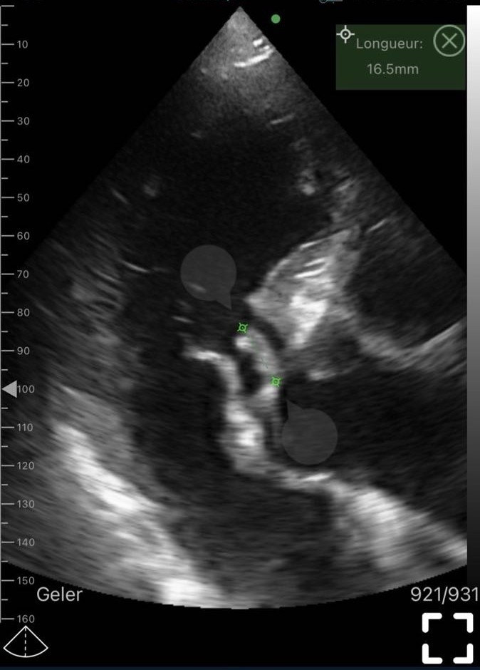
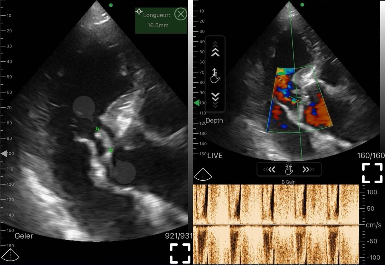
Despite the absence of symptoms, a physician performed echocardiography using a portable ultrasound scanner with a wireless ultrasound probe Wi-Fi, connected to an ultrasound for smartphone system.
Unexpected findings included:
- Aortic valve vegetation
- Severe aortic regurgitation
This case highlights the importance of incorporating portable ultrasound systems into routine preventive evaluation, even in low-risk-appearing patients.
Clinical Interpretation
This patient represents a classic example of clinically silent but structurally advanced cardiac disease.
Key clinical principles illustrated:
- Severe structural heart disease may remain asymptomatic for years
- Physiological compensation can mask symptoms
- Physical examination alone is insufficient for early detection
A portable ultrasound scanner provides immediate visualization of cardiac structures, allowing early identification of pathology before clinical deterioration occurs.
This is especially relevant in point-of-care ultrasound practice, where rapid diagnostic decisions are required.
Examination Methodology
The echocardiographic assessment was performed using a portable ultrasound scanner equipped with a wireless ultrasound probe Wi-Fi, operating via an ultrasound for smartphone interface.
Imaging modalities:
- B-mode – structural assessment of valves and chambers
- Color Doppler – evaluation of regurgitant flow
- PW Doppler – localized flow analysis
- CW Doppler – high-velocity jet assessment
These modalities demonstrate that modern portable ultrasound systems are capable of delivering clinically meaningful cardiac imaging in real-world settings.
Detailed Echocardiographic Findings
- Image Quality
The portable ultrasound scanner provided sufficient spatial and temporal resolution to evaluate:
- Valve leaflet morphology
- Intracardiac structures
- Hemodynamic flow patterns
Modern wireless ultrasound probe Wi-Fi devices are capable of producing clinically reliable echocardiographic images in experienced hands.
- Aortic Valve Morphology
B-mode imaging revealed:
- Thickened aortic valve leaflets
- Irregular leaflet contours
- Impaired coaptation
- Asymmetric closure pattern
These findings are consistent with structural degeneration of the aortic valve apparatus.
- Aortic Valve Vegetation
A mobile hyperechoic mass measuring approximately 16.5 mm was identified attached to the aortic valve.
Key echocardiographic characteristics:
- Irregular, heterogeneous internal structure
- High mobility during cardiac cycle
- Clear attachment to valve leaflet
- Pendulum-like oscillation in blood flow
This pattern is highly suggestive of vegetation, typically associated with infective or inflammatory processes.
Differential diagnostic comparison:
Structure | Mobility | Echogenicity | Attachment |
Vegetation | High | Heterogeneous | Valve leaflet |
Thrombus | Low | Variable | Non-valvular |
Calcification | None | High | Fixed |
Fibrosis | Limited | Moderate | Diffuse |
- Doppler Evaluation of Aortic Regurgitation
Color Doppler demonstrated:
- Wide regurgitant jet originating from the aortic valve
- Turbulent diastolic flow pattern
- Central jet penetration into the left ventricle
CW Doppler confirmed:
- High-velocity regurgitant flow
- Prolonged diastolic phase
- Hemodynamically significant severity
- Hemodynamic Consequences
The following functional changes were observed:
- Left ventricular volume overload
- Progressive chamber dilation
- Preserved systolic function
- Early signs of myocardial remodeling
These findings are consistent with chronic severe aortic regurgitation.
Pathophysiological Analysis
Aortic Valve Vegetation
Potential etiologies include:
- Infective endocarditis
- Subclinical bacterial infection
- Post-inflammatory valvular damage
Clinical risks:
- Systemic embolization
- Structural valve destruction
- Rapid progression of regurgitation
Aortic Regurgitation
Pathophysiological consequences include:
- Chronic volume overload
- Eccentric left ventricular hypertrophy
- Progressive ventricular dilation
- Gradual development of heart failure
Differential Diagnosis
Conditions that must be excluded:
- Degenerative calcific valve disease
- Fibrotic thickening
- Thrombotic masses
- Intracardiac tumors
A portable ultrasound scanner, combined with Doppler imaging, enables accurate differentiation between these conditions based on mobility, structure, and flow characteristics.
Can a portable ultrasound scanner detect aortic valve vegetation in asymptomatic patients?
Yes. A portable ultrasound scanner can detect aortic valve vegetation even in patients without symptoms by identifying a mobile echogenic mass attached to the valve leaflet. In this case, the vegetation was discovered incidentally during routine preventive screening using a wireless ultrasound probe Wi-Fi, highlighting the importance of early echocardiographic evaluation.
How does ultrasound for smartphone help in diagnosing valvular heart disease?
Ultrasound for smartphone enables real-time visualization of cardiac structures, including valve morphology and blood flow patterns. In valvular heart disease, it allows clinicians to detect abnormalities such as leaflet thickening, impaired coaptation, and regurgitant jets using Doppler imaging.
Can severe aortic regurgitation exist without symptoms?
Yes. Severe aortic regurgitation can remain asymptomatic for a long period due to compensatory mechanisms of the left ventricle. Patients may maintain normal daily activity despite significant volume overload, as seen in this clinical case detected by a portable ultrasound scanner.
What makes a wireless ultrasound probe Wi-Fi useful in cardiology?
A wireless ultrasound probe Wi-Fi provides mobility and rapid deployment in clinical settings. It allows immediate bedside imaging, which is essential in detecting acute or chronic cardiac abnormalities such as vegetation and regurgitation without delay associated with traditional echocardiography systems.
How is aortic valve vegetation differentiated from thrombus or calcification?
Differentiation is based on echocardiographic features:
- Vegetation: mobile, irregular, heterogeneous
- Thrombus: less mobile, more uniform
- Calcification: highly echogenic, fixed structure
A portable ultrasound scanner combined with Doppler imaging allows accurate differentiation based on mobility and attachment.
Why was this cardiac pathology not detected earlier in the patient?
In many cases, structural heart disease progresses silently. This patient had no symptoms due to long-term physiological compensation. Only preventive imaging with a portable ultrasound scanner revealed the underlying pathology during routine examination.
What is the role of portable ultrasound systems in preventive cardiology?
Portable ultrasound systems play a key role in preventive cardiology by enabling early detection of structural abnormalities before symptom onset. They support routine screening, especially in high-risk or elderly patients, improving early intervention rates.
Can handheld ultrasound replace traditional echocardiography?
No, but it complements it. Handheld ultrasound devices are highly effective for screening and initial assessment, but comprehensive evaluation may still require full echocardiography systems for detailed measurements and advanced diagnostics.
How accurate is cardiac ultrasound imaging with portable devices?
Modern cardiac ultrasound imaging using portable devices has high diagnostic accuracy when performed by trained operators. While image resolution may be slightly lower than high-end systems, it is sufficient for detecting major structural and functional abnormalities.
What are the clinical risks of untreated aortic valve vegetation?
If untreated, aortic valve vegetation may lead to:
- systemic embolization
- valve destruction
- worsening regurgitation
- potential heart failure
Early detection using a portable ultrasound scanner is therefore critical for prognosis.
Why is point-of-care ultrasound important in cases like this?
Point-of-care ultrasound allows immediate bedside diagnosis without waiting for scheduled imaging. In this case, it enabled instant identification of severe pathology in an asymptomatic patient, significantly accelerating clinical decision-making.
Frequently Asked Questions
What is the most reliable method for PFO diagnosis?
Contrast echocardiography with Valsalva maneuver is the gold standard for detecting PFO and right-to-left shunts.
Can portable ultrasound accurately detect PFO?
Yes, modern portable ultrasound devices provide sufficient image quality and Doppler functionality for reliable PFO diagnosis.
Is portable ultrasound suitable for cardiology use?
Yes, it is widely adopted for structural and functional cardiac assessment, as well as hemodynamic evaluation.
Can Wi-Fi ultrasound devices connect to smartphones or tablets?
Yes, they enable real-time imaging and flexible bedside diagnostics.
Is portable ultrasound effective in emergency situations?
Yes, portable ultrasound allows rapid bedside assessment, aiding immediate clinical decision-making.
Who should use portable ultrasound devices?
They are used by cardiologists, neurologists, emergency physicians, family doctors, and other healthcare professionals.