
Meta Title
Wi-Fi Ultrasound Probe in Ascending Aortic Aneurysm Detection | Extended Clinical Case POCUS
Meta Description
Extended clinical case demonstrating detection of ascending aortic aneurysm using Wi-Fi ultrasound probe and smartphone ultrasound in primary care point-of-care diagnostics.
Introduction
Ascending aortic aneurysm is a progressive and potentially fatal cardiovascular condition characterized by pathological dilation of the ascending segment of the aorta. The disease is often clinically silent until life-threatening complications occur, including acute aortic dissection, rupture, or severe aortic regurgitation.
Early detection is therefore a critical objective in cardiovascular medicine. Traditional diagnostic pathways rely on hospital-based echocardiography, CT angiography, or MRI. However, modern clinical practice increasingly incorporates point-of-care ultrasound (POCUS) as a first-line diagnostic tool.
The development of Wi-Fi ultrasound probes (handheld ultrasound devices connected to smartphones) has significantly expanded the accessibility of vascular imaging. These systems allow clinicians to perform real-time ultrasound examinations directly at the patient’s bedside, in outpatient clinics, emergency settings, and even remote environments.
This extended clinical case demonstrates how a smartphone ultrasound system (ultrasound for phone, portable ultrasound scanner, mini echograph) enabled immediate detection of a clinically significant ascending aortic aneurysm during a routine outpatient evaluation.
The case also highlights the role of portable ultrasound technologies in transforming primary care diagnostics, reducing delays, and improving clinical decision-making efficiency.
Clinical Case Presentation
A 67-year-old male patient was evaluated in an outpatient primary care setting due to persistent but non-specific cardiovascular complaints.
Presenting symptoms
- intermittent dull chest discomfort without radiation to the arms or jaw
- episodic palpitations occurring at rest and during mild exertion
- subjective sensation of internal pulsation in the chest
- general fatigue and reduced exercise tolerance
Although symptoms were non-specific, their persistence raised clinical suspicion of underlying cardiovascular pathology.
Medical history
- arterial hypertension diagnosed more than 15 years earlier
- poor long-term blood pressure control with frequent hypertensive spikes (160–180 mmHg)
- suspected generalized atherosclerotic disease
- sedentary lifestyle with minimal physical activity
- increased cardiovascular risk due to age and comorbidities
Physical examination
- accentuated second heart sound over the aortic valve area
- no signs of acute decompensated heart failure
- stable hemodynamic status at rest
- absence of overt murmurs but presence of subtle vascular suspicion
Given the combination of risk factors and clinical presentation, immediate bedside imaging was indicated.
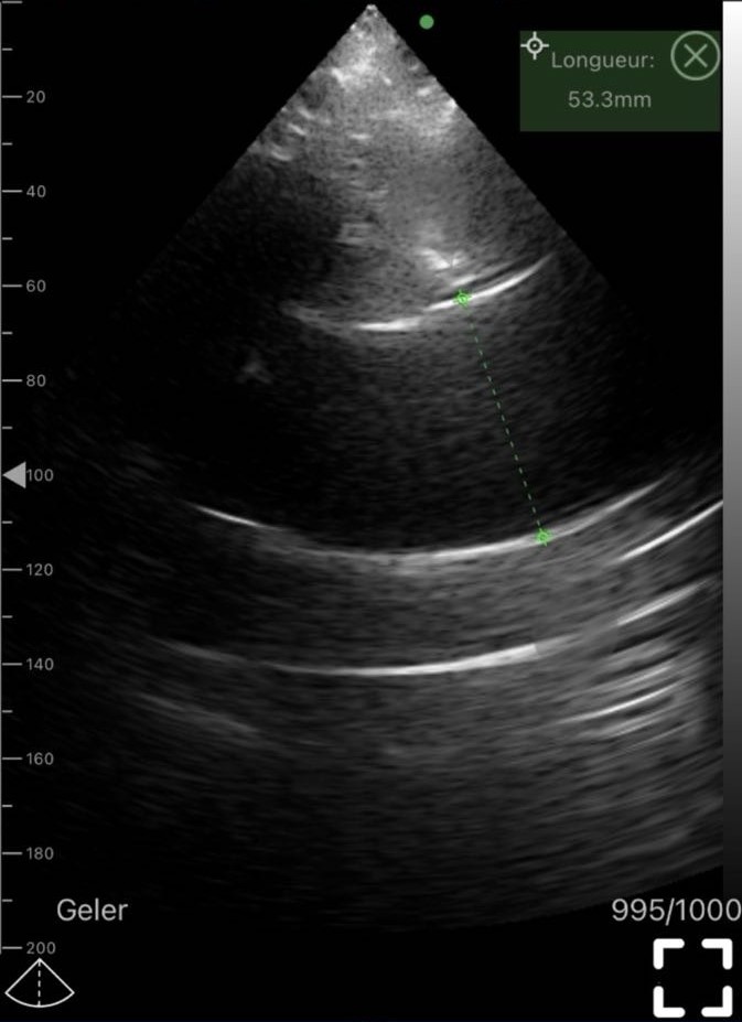
A Wi-Fi ultrasound probe connected to a smartphone-based imaging system was used for point-of-care evaluation. The device was a portable echographic solution provided by Sante Integrale, functioning as a handheld ultrasound scanner designed for real-time cardiovascular assessment.
Point-of-Care Ultrasound Methodology
The examination was performed using a wireless ultrasound probe linked to a mobile ultrasound application.
Scanning protocol
Standard transthoracic echocardiographic views were obtained:
- parasternal long-axis view
- parasternal short-axis view at aortic level
- suprasternal notch view for aortic arch visualization
- limited subcostal assessment for complementary hemodynamic evaluation
The suprasternal view played a key role in accurately visualizing the ascending aorta and identifying abnormal dilation.
Technical characteristics of Wi-Fi ultrasound probe
The handheld ultrasound system provided:
- real-time wireless transmission of ultrasound images to smartphone
- high-resolution sector imaging suitable for cardiac structures
- sufficient penetration depth (approximately 18–20 cm)
- integrated measurement tools with digital calipers
- immediate image storage and clinical documentation capabilities
- portability allowing bedside examination without additional infrastructure
This configuration effectively replaces the need for immediate hospital-based echocardiography in urgent diagnostic triage situations.
Ultrasound Findings
The imaging study revealed significant pathological changes involving the ascending aorta:
- pronounced dilation of the ascending aorta
- measured diameter: 53.3 mm
- marked enlargement exceeding physiological norms (approx. 30–40 mm depending on body size and sex)
- thickened aortic wall with reduced elastic compliance
- absence of visible intimal flap or signs of acute dissection at the time of examination
- no pericardial effusion detected
Clinical interpretation of findings
An ascending aortic diameter above 50 mm is considered a high-risk finding associated with:
- significantly increased risk of spontaneous aortic rupture
- elevated risk of acute aortic dissection
- indication for urgent referral to cardiothoracic surgery evaluation
- need for strict blood pressure control and monitoring
The quality of imaging obtained using a Wi-Fi ultrasound probe connected to a smartphone system was sufficient to perform clinically relevant structural assessment and measurement of the ascending aorta.
Clinical Decision-Making and Significance
The most important aspect of this case is the reduction in diagnostic delay.
Without point-of-care ultrasound, the patient would have required:
- referral to cardiology
- scheduled echocardiography or CT angiography
- waiting period before definitive diagnosis
Instead, immediate bedside imaging allowed:
- real-time detection of life-threatening pathology
- confirmation of clinical suspicion during the same visit
- urgent referral to specialized cardiovascular care
This demonstrates the transformative role of handheld ultrasound devices in primary care and emergency medicine.
Technology Overview: Wi-Fi Ultrasound Probes in Modern Clinical Practice
A Wi-Fi ultrasound probe (portable ultrasound scanner, mini echograph, handheld ultrasound device) is a compact imaging system that connects wirelessly to mobile devices and enables real-time diagnostic imaging.
Core diagnostic capabilities
- cardiac ultrasound (basic echocardiography)
- vascular imaging and aneurysm detection
- abdominal organ assessment
- emergency FAST protocol support
- musculoskeletal and soft tissue imaging
Clinical importance
These devices bridge the gap between physical examination and full radiological imaging, allowing clinicians to extend diagnostic capabilities directly at the point of care.
They are particularly valuable in environments where access to hospital imaging is limited or delayed.
Clinical Applications of Smartphone Ultrasound Systems
- Cardiology
- detection and measurement of aortic aneurysms
- assessment of ventricular size and function
- identification of pericardial effusion
- gross valvular pathology screening
- Emergency medicine
- trauma assessment (FAST protocol)
- internal bleeding detection
- rapid cardiac activity evaluation
- shock state evaluation
- Family medicine
- first-line diagnostic imaging
- chronic cardiovascular disease monitoring
- risk stratification in hypertensive patients
- early detection of vascular pathology
- Vascular diagnostics
- aneurysm screening
- vessel diameter measurement
- peripheral vascular evaluation
- atherosclerotic changes assessment
Diagnostic Accuracy and Clinical Value
When used by trained clinicians, a Wi-Fi ultrasound probe provides:
- high sensitivity for detecting structural cardiovascular abnormalities
- reliable measurement of large vessel diameters
- adequate image resolution for screening-level echocardiography
- immediate clinical feedback for decision-making
In this case, the measured diameter of 53.3 mm was clinically decisive and required urgent escalation of care.
Smartphone-Based Ultrasound in Modern Medicine
The integration of ultrasound imaging into smartphones (ultrasound for phone systems) represents a major shift toward decentralized, accessible diagnostics.
Advantages
- immediate bedside imaging without delays
- reduced dependency on hospital imaging departments
- improved workflow efficiency in outpatient care
- increased accessibility in rural or resource-limited settings
- enhanced clinical decision-making speed
This approach is aligned with modern point-of-care ultrasound (POCUS) medicine, where imaging is integrated into routine clinical examination.
Extended Clinical Applications
Beyond cardiology, portable ultrasound is utilized in multiple medical specialties:
- Emergency medicine: rapid assessment of cardiac and vascular structures
- Neurology: evaluation of patients with TIA or stroke
- Intensive care: hemodynamic monitoring and bedside evaluation
- Family medicine: point-of-care ultrasound for comprehensive assessment
- Internal medicine: multi-organ and systemic evaluations
This versatility underscores the value of portable ultrasound in contemporary clinical practice.
Role of Sante Integrale in Portable Ultrasound Solutions
Reliable and clinically validated equipment is essential for accurate diagnostics. Portable ultrasound systems provided by Sante Integrale meet modern clinical standards and support a wide range of applications. Their devices are used in cardiology, emergency medicine, and family practice, providing clinicians with timely and reliable diagnostic data.
Healthcare providers can see our ultrasound equipment catalog to explore available systems designed for diverse clinical requirements. Sante Integrale ensures equipment integration into clinical workflows without compromising diagnostic quality.
Limitations of Wi-Fi Ultrasound Technology
Despite significant advantages, several limitations must be acknowledged:
- operator-dependent image acquisition and interpretation
- requirement for structured ultrasound training and experience
- limited advanced Doppler functionality in basic handheld models
- not a substitute for comprehensive echocardiography or CT imaging in complex cases
However, for initial detection, screening, and urgent triage, its clinical value is extremely high.
Role of Sante Integrale in Portable Ultrasound Solutions
Sante Integrale provides advanced portable ultrasound systems, Wi-Fi ultrasound probes, and mini echograph devices designed for modern clinical practice.
These systems are widely used in:
- family medicine and outpatient clinics
- cardiology and vascular diagnostics
- emergency and acute care settings
- mobile and decentralized healthcare environments
The availability of such technology enables clinicians to perform real-time diagnostic imaging at the point of care, significantly improving diagnostic speed, accuracy, and patient management efficiency.
Conclusion
This extended clinical case of a 53.3 mm ascending aortic aneurysm in a high-risk hypertensive patient demonstrates the increasing importance of Wi-Fi ultrasound probes and smartphone-based ultrasound systems in modern medicine.
The use of a portable ultrasound scanner enabled:
- immediate visualization of the ascending aorta
- accurate quantification of aneurysm severity
- rapid clinical decision-making and urgent referral
Point-of-care ultrasound is no longer an auxiliary diagnostic method but an essential extension of physical examination.
The integration of handheld ultrasound devices (mini echograph, portable ultrasound scanner, Wi-Fi ultrasound probe) into routine medical practice represents a paradigm shift toward faster, more accurate, and more accessible diagnostics.
Frequently Asked Questions
Can a Wi-Fi ultrasound probe detect an ascending aortic aneurysm?
Yes. It can clearly visualize and measure the ascending aorta in real time, as demonstrated in this clinical case.
Is smartphone ultrasound reliable in clinical practice?
Yes, for screening and initial diagnostic evaluation when performed by trained clinicians.
Can handheld ultrasound replace hospital imaging?
Handheld ultrasound systems, including advanced portable devices such as those provided by Sante Integrale, can deliver high-quality imaging and in many clinical situations perform a fully sufficient diagnostic role at the point of care. However, comprehensive hospital imaging modalities (such as full echocardiography protocols, CT, or MRI) may still be required for detailed assessment, complex cases, or pre-surgical planning.
Who should use portable ultrasound devices?
Trained physicians in cardiology, family medicine, and emergency care.
What is the main advantage of Wi-Fi ultrasound technology?
Immediate bedside diagnosis without waiting for imaging referrals.