
Introduction
Portable ultrasound has become a cornerstone in modern cardiology, offering clinicians the ability to perform high-quality echocardiography at the bedside or in a variety of clinical settings. One of the clinically significant applications of portable ultrasound is the diagnosis of patent foramen ovale (PFO), a congenital cardiac anomaly found in approximately 25% of adults. While often asymptomatic, PFO can contribute to cryptogenic stroke or transient ischemic attack (TIA), making its timely detection crucial.
Accurate PFO diagnosis requires a structured echocardiographic approach, including baseline transthoracic imaging, contrast studies, and functional maneuvers such as the Valsalva. In the past, these evaluations were largely restricted to stationary ultrasound systems. The advent of portable, wireless Wi-Fi ultrasound devices has transformed this landscape, allowing clinicians to perform comprehensive and reliable imaging in outpatient clinics, emergency rooms, intensive care units, and even remote or pre-hospital settings.
This article presents a detailed clinical case of PFO diagnosis in an adult patient using a portable ultrasound system. It provides an in-depth discussion of echocardiographic methodology, key ultrasound findings, diagnostic accuracy, and practical implications for clinical decision-making. Furthermore, it highlights the advantages of modern portable Wi-Fi ultrasound solutions and their integration into everyday clinical practice, including the use of systems provided by Sante Integrale, a trusted supplier of portable diagnostic equipment.
Clinical Case: Transient Ischemic Attack and the Need for PFO Evaluation
A 52-year-old male patient presented with neurological symptoms consistent with a TIA, including transient unilateral weakness and speech disturbances. TIA is characterized by temporary disruption of cerebral blood flow, usually resolving within 24 hours, and serves as a warning for potential stroke.
The primary clinical objective in this case was to identify the source of embolism. Initial workup, including Doppler ultrasonography of extracranial arteries, excluded significant stenosis. Consequently, a cardioembolic mechanism was suspected. Paradoxical embolism, in which a venous thrombus passes into arterial circulation through an intracardiac shunt such as a PFO, was considered a likely cause.
The patient was referred for echocardiographic evaluation, emphasizing the detection of PFO. A portable Wi-Fi ultrasound system was employed to provide immediate bedside assessment with high-resolution imaging.
Echocardiographic Findings Using Portable Ultrasound
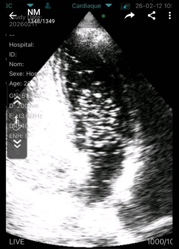
The examination was conducted using a portable ultrasound system integrated with a mobile device, providing real-time imaging and enabling comprehensive assessment of cardiac structure and function.
Key findings included:
- Normal dimensions of cardiac chambers
- Intact valvular structures with no regurgitation or stenosis
- Preserved left ventricular systolic function
- Presence of atrial septal aneurysm
- Positive contrast echocardiography indicating right-to-left shunt
Contrast echocardiography with microbubbles during the Valsalva maneuver revealed clear passage of bubbles from the right atrium to the left atrium, confirming the diagnosis of PFO.
These findings established a causal relationship between the patient’s TIA and intracardiac shunting, supporting a clinical decision for transcatheter closure.
Clinical Rationale for PFO Diagnosis
PFO diagnosis is critical in patients with unexplained ischemic events. When vascular causes are excluded, attention must focus on potential cardiac sources. A PFO can provide a pathway for paradoxical embolism, especially in the presence of an atrial septal aneurysm, which increases shunt mobility and the risk of thrombus passage.
Contrast-enhanced echocardiography remains the reference method for detecting right-to-left shunts. Incorporating portable ultrasound technology allows clinicians to perform these assessments in a broader range of clinical settings, improving the speed and efficiency of diagnosis.
Examination Technique and Technical Capabilities of Portable Ultrasound
Portable Wi-Fi ultrasound systems support comprehensive echocardiographic examinations.
Technical capabilities:
- High-resolution B-mode imaging for structural assessment
- Color Doppler for visualization of blood flow
- Pulsed-wave Doppler for hemodynamic evaluation
- Real-time image transmission via Wi-Fi to smartphones, tablets, or laptops
- Full integration with mobile devices for bedside diagnostics
These capabilities allow clinicians to perform echocardiography with the same diagnostic quality as stationary systems while benefiting from mobility and rapid deployment.
Healthcare providers can learn more about portable ultrasound systems to explore advanced diagnostic workflows and clinical integration.
Ultrasound Features of PFO
Identifying a PFO requires careful observation of structural and functional signs.
Key echocardiographic indicators:
- Atrial septal aneurysm with abnormal mobility
- Microbubble passage across the interatrial septum during contrast study
- Right-to-left shunt visualization during Valsalva maneuver
- Clear delineation of interatrial communication
Recognizing these features is essential for accurate diagnosis and informs clinical decisions regarding closure or monitoring.
Diagnostic Accuracy and Clinical Reliability
Modern portable ultrasound devices have demonstrated high reliability and diagnostic accuracy.
Clinical capabilities:
- Detailed structural visualization using B-mode
- Reliable detection of intracardiac shunts using color and pulsed Doppler
- Accurate hemodynamic assessment comparable to stationary systems
These features ensure portable ultrasound can serve as a primary diagnostic tool in routine and emergency cardiology practice.
Healthcare professionals who explore wireless ultrasound solutions appreciate their ability to deliver consistent, high-quality imaging across various clinical environments.
Advantages of Portable Ultrasound in Clinical Practice
Portable ultrasound provides multiple practical advantages:
Key benefits:
- Mobility: deployable in hospitals, clinics, and pre-hospital settings
- Speed: immediate bedside diagnostics without patient transport
- Efficiency: streamlined workflow for rapid decision-making
- Accessibility: available for a broad range of specialties
- Flexibility: adaptable to diverse patient populations and clinical needs
These advantages are particularly important for emergency medicine, cardiology, and neurology, where rapid diagnosis is critical.
Extended Clinical Applications
Beyond cardiology, portable ultrasound is utilized in multiple medical specialties:
- Emergency medicine: rapid assessment of cardiac and vascular structures
- Neurology: evaluation of patients with TIA or stroke
- Intensive care: hemodynamic monitoring and bedside evaluation
- Family medicine: point-of-care ultrasound for comprehensive assessment
- Internal medicine: multi-organ and systemic evaluations
This versatility underscores the value of portable ultrasound in contemporary clinical practice.
Role of Sante Integrale in Portable Ultrasound Solutions
Reliable and clinically validated equipment is essential for accurate diagnostics. Portable ultrasound systems provided by Sante Integrale meet modern clinical standards and support a wide range of applications. Their devices are used in cardiology, emergency medicine, and family practice, providing clinicians with timely and reliable diagnostic data.
Healthcare providers can see our ultrasound equipment catalog to explore available systems designed for diverse clinical requirements. Sante Integrale ensures equipment integration into clinical workflows without compromising diagnostic quality.
Comparison: Portable vs Traditional Ultrasound Systems
Feature | Portable Ultrasound | Traditional Systems |
Mobility | High | Limited |
Image quality | Very high | Very high |
Setup time | Minimal | Longer |
Accessibility | Wide | Restricted |
Flexibility | High | Moderate |
Portable ultrasound complements traditional systems and enhances flexibility in diagnostics.
Clinical Impact on Patient Management
Integrating portable ultrasound into practice improves patient care.
Key outcomes:
- Faster diagnosis
- Earlier intervention decisions
- Reduced need for patient transfer
- Improved workflow efficiency
- Enhanced patient safety
In the presented case, timely PFO diagnosis enabled appropriate interventional management, reducing the risk of recurrent ischemic events.
Conclusion
Portable ultrasound is a reliable, high-quality diagnostic tool for PFO in adults. It enables accurate echocardiographic assessment, identification of intracardiac shunts, and informed clinical decision-making.
Modern Wi-Fi-enabled systems provide diagnostic precision, mobility, and real-time imaging, supporting evidence-based interventions in cardiology and other specialties.
Solutions available from Sante Integrale allow clinicians to integrate portable ultrasound into diverse clinical workflows effectively, enhancing diagnostic efficiency and patient outcomes.
Frequently Asked Questions
What is the most reliable method for PFO diagnosis?
Contrast echocardiography with Valsalva maneuver is the gold standard for detecting PFO and right-to-left shunts.
Can portable ultrasound accurately detect PFO?
Yes, modern portable ultrasound devices provide sufficient image quality and Doppler functionality for reliable PFO diagnosis.
Is portable ultrasound suitable for cardiology use?
Yes, it is widely adopted for structural and functional cardiac assessment, as well as hemodynamic evaluation.
Can Wi-Fi ultrasound devices connect to smartphones or tablets?
Yes, they enable real-time imaging and flexible bedside diagnostics.
Is portable ultrasound effective in emergency situations?
Yes, portable ultrasound allows rapid bedside assessment, aiding immediate clinical decision-making.
Who should use portable ultrasound devices?
They are used by cardiologists, neurologists, emergency physicians, family doctors, and other healthcare professionals.